Ir al contenido principal
Publicación

ESPECIAL EN INGLÉS. Científica Dental vol 17 2020

Año: 2020
Número: 4
Volumen: 17

Original article

Published in spanish Científica Dental Vol.17. nº1. 2020. www.cientificadental.es

Radiographic diagnosis of impacted maxillary canines: Comparison between two and three dimensions

Resumen

Introduction: An impacted canine is a very common condition and raises several clinical complications. Early and exact diagnosis is important in order to minimise the risks and subsequent complications. The objective of this study is to analyse the effectiveness of two dimensions in the volumetric diagnosis for impacted maxillary canines, using the lines proposed by Alqerban as a reference.

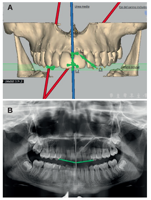
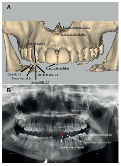
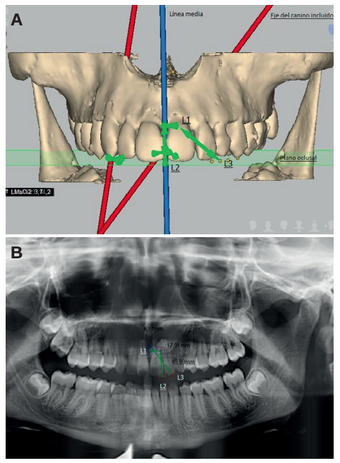
Methods: An orthodontic study of the maxilla using orthopantomography with cone beam computed tomography (CBCT) at the Madrid European University Clinic was performed on 27 patients selected with 36 maxillary impacted canines. Three reference lines were drawn based on the distance from the cusp of the canine to the occlusal plane (L1), to the midline (L2) and to its ideal eruption site (L3), in both the orthopanthomography and the CBCT. As ideal reference values, we selected a control group of 36 erupted maxillary canines.

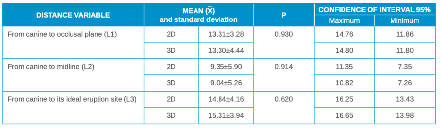
Results: The results were compared in 2 and 3 dimensions using the Student’s t test, after verifying their normal distribution using the Anderson-Darling contrast test. Statistical significance (p > 0.05) was not obtained for any of the variables studied.

Conclusions: The use of CBCT is vital to ensure good diagnosis of the canine position and its relationship with adjacent structures and thus establish an adequate treatment plan. However, orthopantomography provides sufficient information for initial planning.

Abstract
Este artículo está exclusivamente disponible para su descarga en PDF. | 03/29/2024
Palabras clave
Cone beam computed tomography, Dental inclusion, Impacted maxillary canines, Impacted tooth, Orthopantomography
Introducción

Canines are of vital importance in facial and oral aesthetics, as well as in the functionality and development of occlusion. Both Andrews1 with his six keys to occlusion and the latest articles by Clark2 demonstrate the importance of the canine in occlusion. There is no doubt that the canine is one of the pillars in the ideal occlusion scheme proposed by nature. If it is in an aberrant position, it can cause alterations in the entire occlusion system. Due to its anatomy, the maxillary canine guides mandibular movements and supports the forces of occlusion, with a large crown compared to the size of the mandibular tooth itself, and is the tooth with the greatest stability. Its roots are the longest and widest, so these teeth have a firm anchorage in the alveolar bone. Clinically, canines are the teeth that should be lost last. Due to their strategic location in the mouth, they are the cornerstones of the dental arch3 .

The maxillary canine is the permanent tooth with the longest eruption path. It begins forming with a mesial tilt and rapid growth, then slows down as it straightens or even shows a slightly distal diversion.4 This change in speed and inclination corresponds to the contact of the canine with the distal area of the lateral incisor, at approximately 9 years of age. Hence the important role played by the upper lateral incisor in the eruption of the canine. The prevalence and incidence of an impacted maxillary canine is widely reported in the literature. The earliest articles we found in this regard were by Cramer in 19295 and Mead in 19306 . These describe an incidence of 1.4% and 1.57%, respectively, after selecting a sample of American white males. Other authors expand and modify the sample, and obtain prevalences of 0.92% (Dachi7 ), 1.8% (Thilander and Jakobsson8 ), 2.2% (Thilander and Myberg9 ), 3.61% (Aitasa- lo10) and 2.8% (Ericson and Kurol4,11-13).

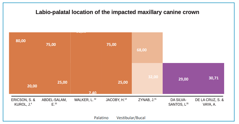
For the interarch position, the classification refers to maxillary canines impacted by the palatal or vestibular. According to this classification14, Jacoby found that 92.31% of patients (a ratio of 12:1) had a palatal impaction, on later expansion of the sample, a ratio of 6.6:1, palatal vs vestibular, was found. Other authors, such as Gaulis and Joho15 obtained a ratio lower than 2:1. The international consensus is for a ratio of 3:1. The current classification, proposed by authors such as Stivaros and Mandall16, reduces the percentage of palatal inclusions to 61%, while vestibular inclusions appear in 5% of patients. For these authors, 34% of canines would be positioned at an intermediate point in the arch. For Rimes et al,17 the proportion of canines impacted palatally is 44%, while those displaced in the vestibular position is 38%. Syrynska18, however, reported 60.3% for palatal canines and 20.6% for vestibular; while 19.2% were in an intermediate position in the alveolus.

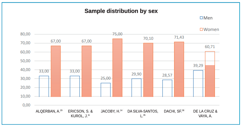
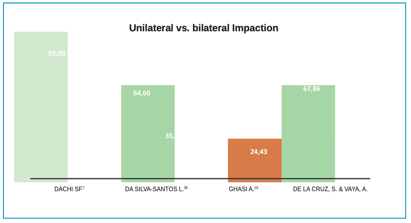
The literature suggests impaction occurs more in women than men. Dachi7 reports 78.57%, Gashi19 77.10% and Bishara20 suggests a ratio of 2:1 for maxillary canine impaction in women over men, which is confirmed by Cooke21.

Regarding bilaterality, 8% of patients have a bilateral impaction according to Dachi7 , Bishara20, Manne22 and Yadav23. Shirazi24, however, found no association in gender for unilateral or bilateral impaction in maxillary canines.

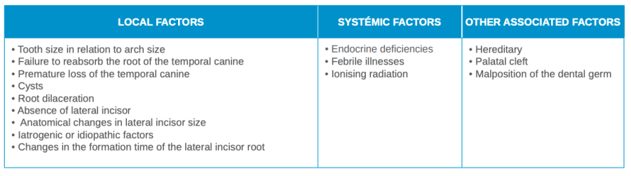
Most authors associate palatal inclusion with the Caucasian race, at 5.9%, while vestibular inclusion is associated with Asians, 1.7%25-27. The impaction ratio of Caucasian patients to African or Asian patients is 2:1, according to Peck and Peck28. Etiological factors associated with impacted canines are shown in Table 1.

Table 1. Factors associated with impacted maxillary canines28.
Bibliografía
1

Andrews L. The six keys to normal occlusion. Am J Orthod 1972; 62 (3): 296-309.

2

Clark J, Evans R. Functional occlusion. A review. J Orthod 2001; 28 (1): 76-81.

3

Kraus B, Jordan R, Abrams L. Anatomía dental y oclusión. : Lippincott Willams and Wilkins 1969.

4

Ericson S, Kurol J. Radiographic of ectopically erupting canines. Am J Orthod Dentofacial Orthop 1987; 91 (6): 483-492.

5

Cramer HC. Dental survey of one thousand adult males: A statistical study correlated with physical and laboratory findings. J Am Dent Assoc 1929; 16: 122.

6

Mead SV. Incidence of impacted teeth. Int J Orthod 1930; 16: 885-890.

7

Dachi SF, Howell FV. A survey of 3.874 routine full-mouth radiographs. A study of impacted teeth. J Oral Maxillofac Surg 1961; 14 (10): 1165-1169.

8

Thilander B, Jakobsson S. Local factors in impaction of maxillary canine. Acta Odontol Scand 1968; 26 (2): 145-168.

9

Thilander B, Myrberg N. The prevalence of malocclusion in Swedish schoolchildren. Scand J Dent Res 1973; 81: 12-21.

10

Aitasalo K, Lehtinen R, Oksala E. An orthopantomographic study of prevalence of impacted teeth. Int J Oral Surg 1972; 1 (3): 117–120.

11

Ericson S, Kurol J. Radiographic assessment of maxillary canine eruption in children with clinical signs of eruption disturbance. Eur J Orthod 1986; 8 (1): 133-140.

12

Ericson S, Kurol J. Early treatment of palatally erupting maxillary canines by extraction of the primary canines. Eur J Orthod 1988; 10 (1): 283-295.

13

Ericson S, Kurol J. Resorption of maxillary lateral incisor caused by ectopc eruption of the canines. A clinical and radiographic analysis of predisposing factors. J Orthod Dentofac Orthop 1988; 4: 503-513.

14

Jacoby H. The “ballista spring” system for impacted teeth. Am J Orthod 1979; 75: 143-151.

15

Gaulis R, Joho JP. Parodonte marginal de canines superieures incluses: Evaluation suite a differentes methodes d’acces chimrgical et de systeme orthodontique. Rev Med Suisse 1978; 88: 1249-1261.

16

Stivaros N, Mandall NA. Radiographic factors affecting the management of impacted upper permanent canines. J Orthod 2000; 27 (2): 169-173.

17

Rimes RJ, Mitchell CN, Willmot DR. Maxillary incisor resorption in relation to the ectopic canine: a review of 26 patients. Eur J of Orthod 1997; 19: 79-84.

18

Syrynska M, Budzynska A. The incidence of uni-and bilateral impacted maxillary canines and their position in dental arch depending on gender and age. Ann Acad Med Stetin 2008; 54 (2): 132-137.

19

Gashi A, Kamberi B. Ademi-Abdyli R. The incidence of impacted maxillary canines in Kosovar population. Int Sch Res Notices. 2014; 1: 1-4.

20

Bishara S. Impacted maxilary canines. Am J Orthod Dentofacial Orthop 1992; 101 (2): 159-171.

21

Cooke J, Wang HL. Canine Impactions: Incidence and Management. Int J Periodontics Restorative Dent 2006; 23 (5): 483-491.

22

Manne R, Gandikota C, Juvvadi SR. Impacted canines: etiology, diagnosis, and orthodontic management. J Pharm Bioallied Sci 2012; 4 (2): 5234-5238.

23

Yadav R, Shrestha BK. Maxillary Impacted Canines: A Clinical Review. Orthod J Nepal 2013; 3 (1): 63-68.

24

Shirazi Z, Kjaer I. Is the Etiology Behind Palatal Unilateral and Palatal Bilateral Maxillary Canine Ectopia Different? Dent Hypotheses 2018; 9 (1): 3-10.

25

Montelius GA. Impacted teeth. A comparative study of Chinese and Caucasian Dentitions. J Dent Res 1932; 12: 931.

26

Oliver RG, Mannion JE, Robinson JM. Morphology of the maxillary lateral incisor in cases of unilateral impaction of the maxillary canine. Br J Orthod 1989; 16: 9-16.

27

Kramer RM, Willams AC. The incidence of impacted teeth. A suvery at Harlem Hospital. Oral Surg Oral Med Oral Pathol Oral Radiol Endod 1970; 29 (2): 237-241.

28

Peck S, Peck L, Kataja M. The palatally displaced canine as a dental anomaly of genetic origin. Angle Orthod 1994; 64: 249-256.

29

Alqerban A. Comparision of two CBCT systems VS panoramic imagin for localization of impacted. Eur J Orthod 2011; 33: 93-110.

30

Ericson S, Kurol J. Radiographic of ectopically erupting canines. Am J Orthod Dentofacial Orthop 1987; 91 (6): 483-492.

31

Walter L. Three-dimensional localization of maxilary canines with CBCT. Am J Orthod Dentofacial Orthop 2005; 128 (4): 418-23.

32

Walker L, Enciso R, Mah J. Three-dimensional localization of maxillary canines with cone-bem computed tomography. Am J Orthod Dentofacial Orthop 2005; 128: 418-423.

33

Abdel-Salam E, El-Badrawy A, Tawfik AM. Multi-detector dental CT in evaluation of impacted maxillary canine. Egypt J of Radiol Nucl Med 2012; 43 (4): 534.

34

Oliver R, Mannion J, Robinson J. Morphology of the maxillary lateral incisor in cases of unilateral impactation of the maxillary canine. Br J Orthod 1989; 16: 9-16.

35

Da Silva-Santos L, Bastos LC, Olveira-Santos C. CBCT findings of impacted upper canines. Imaging Sci Dent 2014; 44: 287-292.

36

Dachi SF, Howell FV. A survey of 3.874 routine full-mouth radiographs. A study of impacted teeth. Oral Surg Oral Med Oral Pathol Oral Radiol Endod 1961; 14 (10): 1165-1169.

37

Stivaros M. Radiographic factors affecting the management of impacted upper permanent canines. J Orthod 2000; 27: 169- 173.

38

Sawamura T, Minowa K, Nakamura M. Impacted teeth in the maxillr: usefulness of 3D Dental-CT for preoperative evaluation. Eur J Radiol 2003; 47: 221-226.

39

Chen Y, Duan P, Chend Y. Three dimensional spiral computed tomographic imging: a new approach to the diagnosis and treatment planning of impacted teeth. Am J Orthod Dentofacial Orthop 2006; 130: 112-116.

40

Ferrario S. Dal 2D al 3D nella diagnosi e pianifiazione terapeuticaa dei canini mascellaai inclusi. Eur J Oral Implantol 2012; 11 (5): 96-107.