Caso clínico
Beca Campoy T, Sánchez-Labrador L, Cortés-Bretón J, Blanco Antona LA, Martínez-González JM. Comportamiento clínico, radiográfico e histomorfométrico del diente autólogo como biomaterial en elevación de seno maxilar de acceso lateral. Caso clínico con seis meses de seguimiento post-carga protésica. Cient. Dent. 2024; 21; 1; 30-39
Comportamiento clínico, radiográfico e histomorfométrico del diente autólogo como biomaterial en elevación de seno maxilar de acceso lateral. Caso clínico con seis meses de seguimiento post-carga protésica
Introducción: la pérdida ósea tras las extracciones puede requerir en el maxilar posterior la realización de una elevación de seno para la correcta colocación de implantes. El hueso autólogo se considera el gold estándar, pero tiene una alta tasa de reabsorción y morbilidad, haciendo que haya otras alternativas como el diente autólogo, con buenos resultados en procedimientos regenerativos. Este caso clínico evalúa a nivel clínico, radiográfico e histomorfométrico el empleo del diente autólogo en elevación de seno maxilar y el comportamiento de dos implantes colocados de manera diferida.
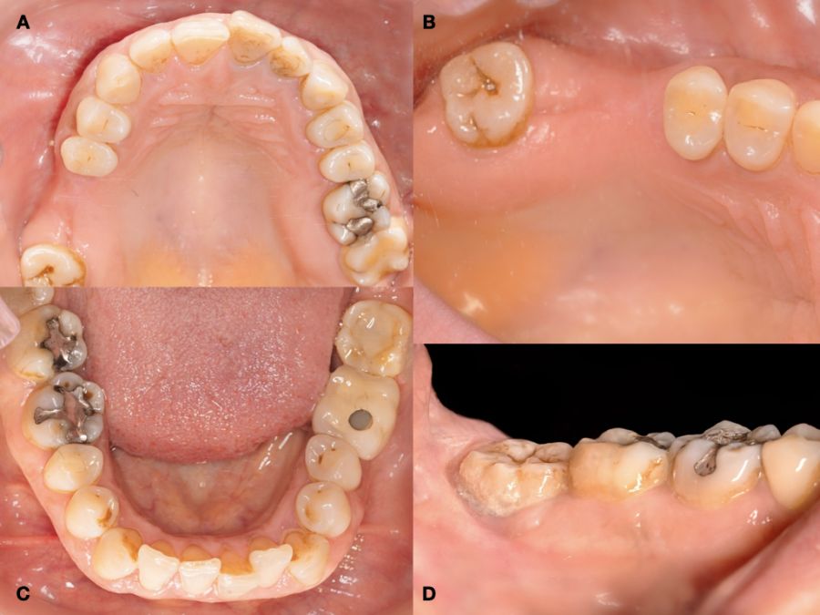
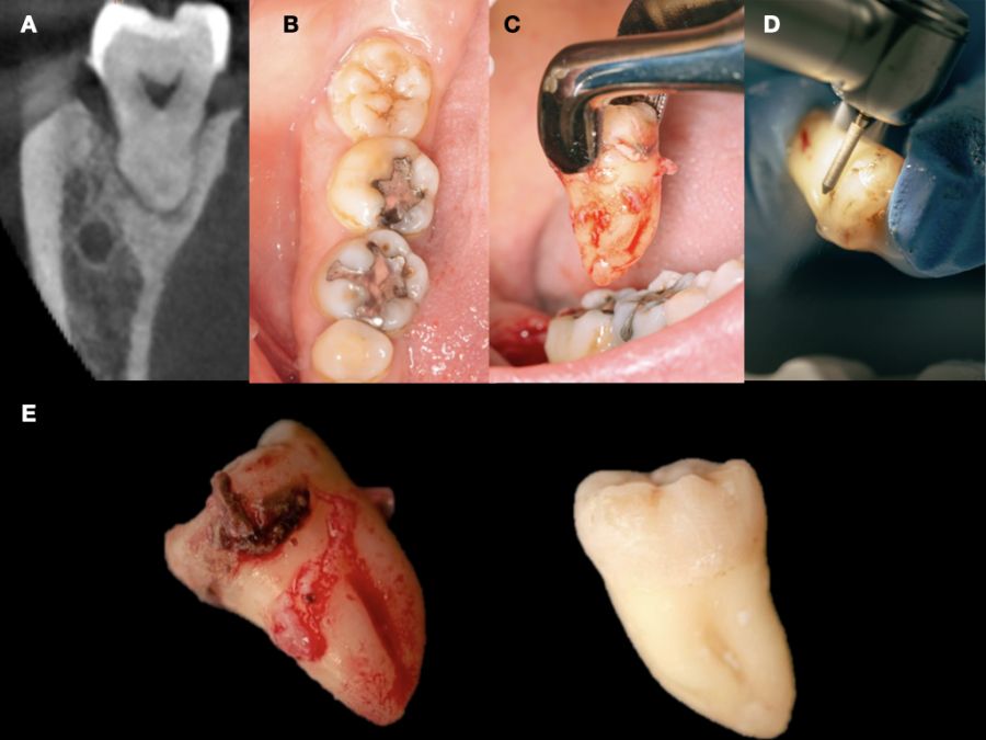
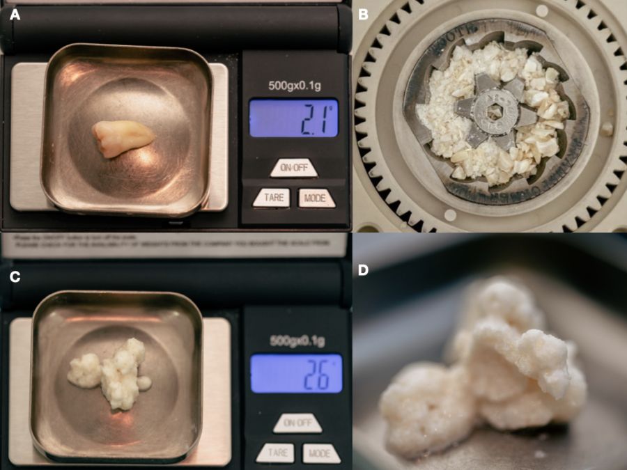
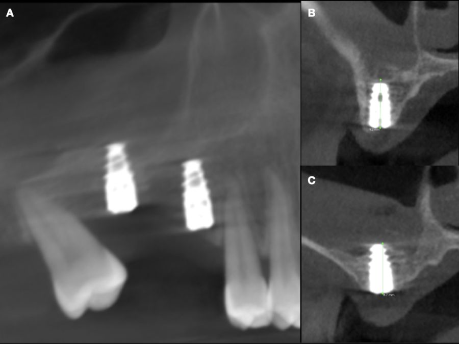
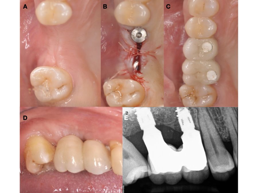
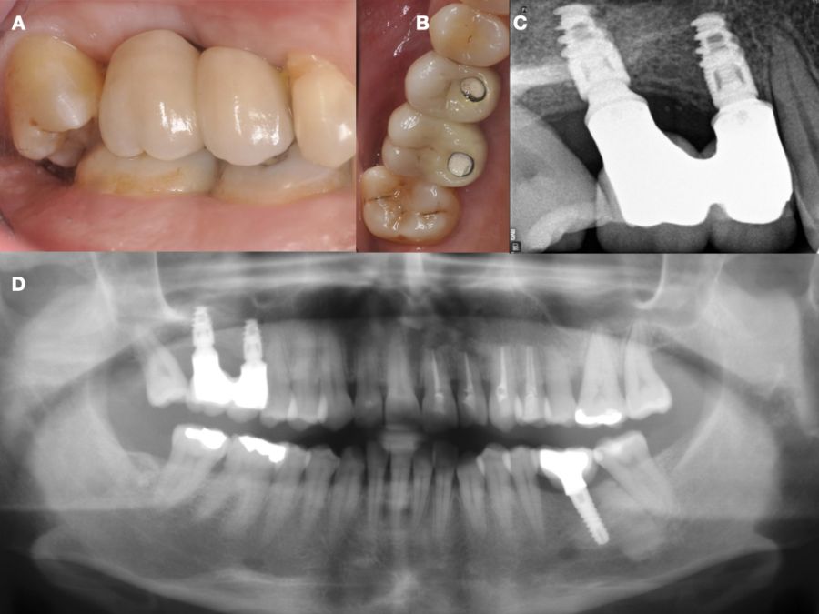
Caso clínico: se presenta el caso de una mujer de 48 años, que acudió a consulta para reponer el sector posterior derecho. Se realizó la exodoncia del 4.8 como diente donante para utilizarlo como biomaterial en una elevación de seno de acceso lateral, colocando dos implantes seis meses tras la intervención, y evaluándolos seis meses tras su carga protésica.
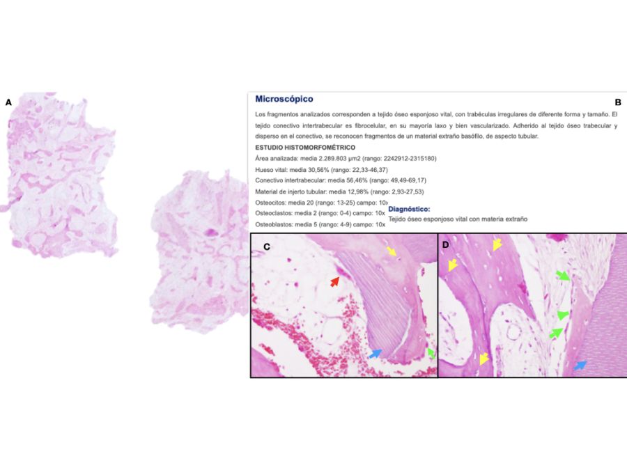
Discusión: el diente autólogo en el presente caso clínico presentó un 30,56% de hueso neoformado tras seis meses de espera, con mejores resultados que al emplear aloinjertos y xenoinjertos. Además, diferentes aspectos culturales y étnicos respaldan la aceptación del diente autólogo por parte de los pacientes, aunque se necesitan más estudios a largo plazo para evaluar la estabilidad de este tipo de injerto en elevación de seno maxilar.
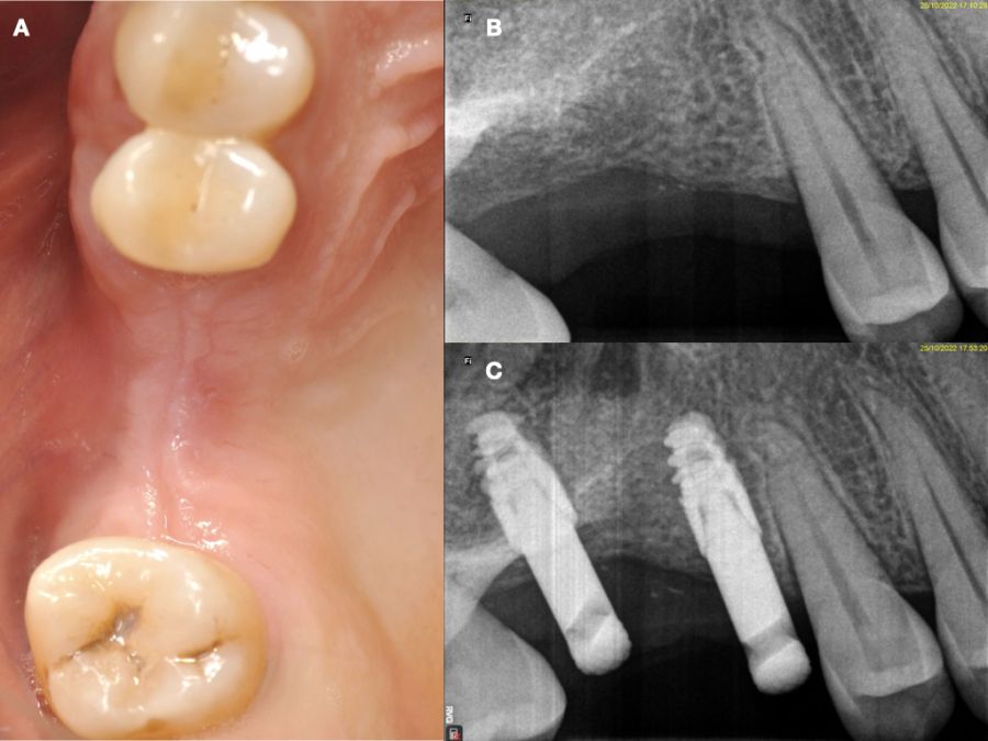
Conclusiones: el diente autólogo en la elevación de seno ofrece biocompatibilidad, baja incidencia de complicaciones y buena aceptación por parte del paciente, con un buen comportamiento clínico y radiográfico de los implantes, a pesar del poco tiempo transcurrido en este caso tras la carga.
Clinical, radiographic and histomorphometric behaviour of the autologous tooth as a biomaterial in lateral access maxillary sinus elevation. Case report with six months of post-prosthetic loading follow-up
Introduction: bone loss after extractions may require a sinus elevation to be performed in the posterior maxilla for the correct placement of implants. Autologous bone is considered the gold standard, but has a high rate of resorption and morbidity, leading to other alternatives such as autologous tooth, with good results in regenerative procedures. This case report evaluates at the clinical, radiographic and histomorphometric level the use of the autologous tooth in maxillary sinus elevation and the behaviour of two implants placed in a delayed manner.
Case report: the case is presented of a 48-year-old woman who came for consultation to replace the right posterior sector. The extraction of 4.8 as a donor tooth was performed to use it as a biomaterial in a lateral access sinus elevation, placing two implants six months after the intervention, and evaluating them six months after their prosthetic loading.
Discussion: the autologous tooth in this case report showed 30.56% of newly formed bone following a six-month wait, with better results than when allografts and xenografts were used. In addition, different cultural and ethnic aspects support the acceptance of the autologous tooth by patients. However, more long-term studies are needed to evaluate the stability of this type of graft in maxillary sinus elevation.
Conclusions: the autologous tooth in the sinus elevation offers biocompatibility, low incidence of complications and good patient acceptance, with good clinical and radiographic behaviour of the implants, despite the short time elapsed in this case after loading.
Keywords: Autologous tooth; Lateral sinus elevation; Autologous dentin.
Tres meses tras una extracción dental, se produce una pérdida del 50% de las dimensiones óseas iniciales del alveolo, cobrando especial importancia en la región posterior del maxilar. En esta región anatómica, la pérdida de los dientes antrales produce una neumatización tridimensional del seno maxilar, pudiendo alcanzar la cresta alveolar y la región anterior, la zona tuberositaria y el hueso cigomático. Este doble proceso de neumatización y remodelación ósea reduce la disponibilidad ósea en sentido horizontal y vertical, pudiendo comprometer el tratamiento con implantes y su estabilidad a largo plazo1-6.
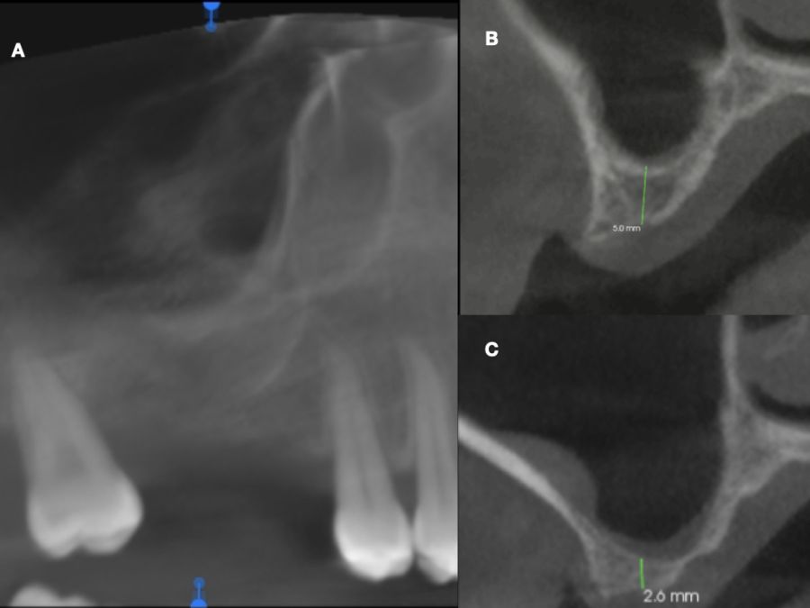
En estos casos, la técnica más predecible para la reconstrucción ósea es la elevación de seno maxilar, que permite una correcta colocación de implantes y su posterior restauración implantosoportada, mejorando la cantidad y calidad ósea del lecho implantado. Entre las técnicas de elevación del seno maxilar, la técnica de acceso lateral está indicada cuando la altura ósea vertical es ≤ 4 mm, colocando los implantes de manera diferida, mientras que con una altura ≥ 5 mm, se recomienda la técnica de elevación de seno transcrestal e implantes más cortos, o técnica abierta con implantes simultáneos de mayor longitud7,8.
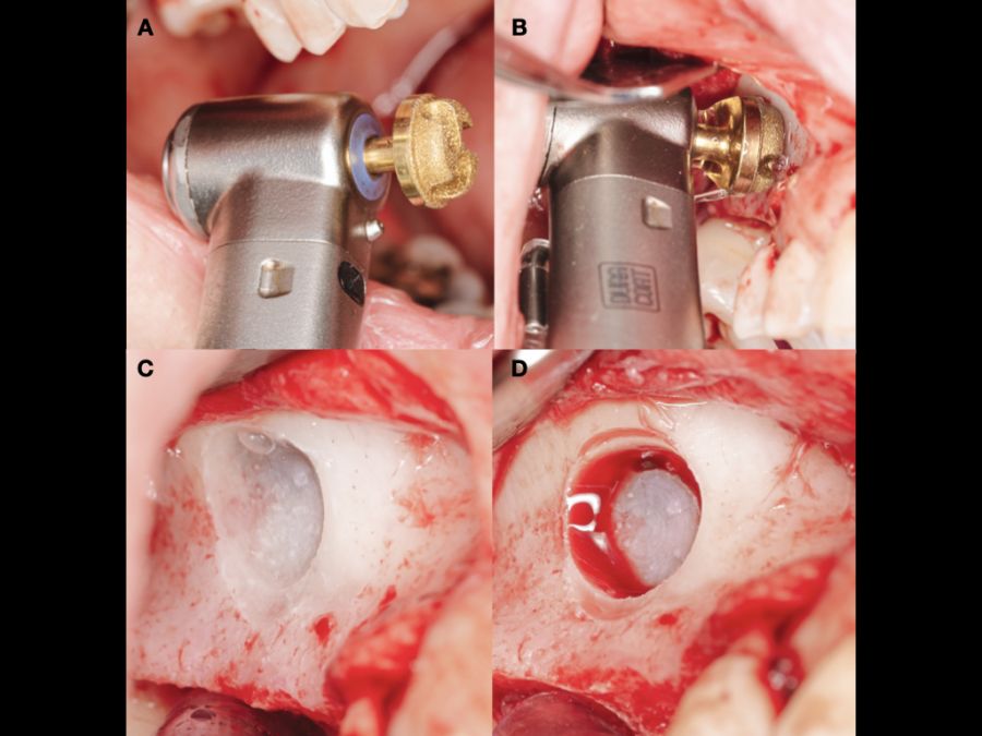
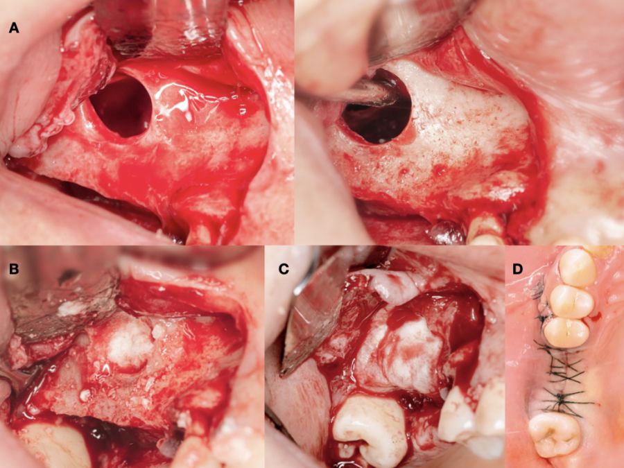
La elevación de seno de acceso lateral, también denominada técnica abierta, es una técnica bien documentada, habiendo sido descrita por Tatum9 en 1976 y publicada posteriormente por Boyne y James10 en 1980. Consiste en la realización de un colgajo a espesor total, para poder acceder a la cara anteroexterna del seno maxilar, y mediante osteotomía realizar una ventana en la cortical vestibular, exponiendo la membrana de Schneider. Una vez expuesta esta membrana, será despegada cuidadosamente y se elevará hasta tener una posición horizontal para formar el nuevo suelo del seno, colocándose posteriormente un biomaterial de relleno. Se puede colocar una membrana reabsorbible o no antes de suturar, para evitar el desplazamiento del injerto y la colonización del interior del seno por el periostio procedente del colgajo11,12.
Entre los biomateriales empleados en esta técnica, el hueso autólogo es actualmente considerado como gold standard debido a que proporciona un buen andamiaje para la osteoconducción, contiene factores de crecimiento para favorecer la osteoinducción y células osteocompetentes para permitir los procesos de osteogénesis, aunque ciertos inconvenientes como la morbilidad de la zona donante, su limitada disponibilidad y su alta tasa de reabsorción, pueden condicionar su uso13,14.
Por estos motivos se han empleado diferentes sustitutos óseos (aloinjertos, xenoinjertos y materiales aloplásticos). La mayoría de estos biomateriales solo presentan propiedades osteoconductoras y tiempos de reabsorción muy variables, que van desde los muy cortos (derivados del ácido poliglicólico y poliláctico) a los muy largos (hidroxiapatitas), mientras que otros pueden presentar reacciones inmunes (aloinjertos). Debido a estas desventajas, se han incrementado en los últimos años los estudios sobre el comportamiento del diente en diferentes procedimientos regenerativos, por su similitud con el hueso humano15,16.
Kim y cols.17describieron las propiedades osteoinductivas y osteoconductoras del diente, así como una menor morbilidad y mayor aceptación por parte del paciente, habiéndose demostrado su buen comportamiento clínico y radiográfico en procedimientos de elevación de seno maxilar, regeneración ósea guiada y preservación alveolar18.
El objetivo del presente caso clínico es evaluar clínica, radiográfica e histomorfométricamente el uso del diente autólogo como biomaterial en elevación de seno maxilar, así como el comportamiento clínico y radiográfico de dos implantes colocados de manera diferida a la elevación de seno, y su evolución seis meses tras la carga protésica de los mismos.
Chappuis V, Araújo MG, Buser D. Clinical relevance of dimensional bone and soft tissue alterations post-extraction in esthetic sites. Periodontol 2000 2017; 73 (1):73–83.
Fischer KR, Gotz W, Kauffmann F, Schmidlin PR, Friedmann A. Ridge preservation of compromised extraction sockets applying a soft cortical membrane: a canine proof-of-principle evaluation. Ann Anat 2020; 231:151524.
Lawson W, Patel ZM, Lin FY. The development and pathologic processes that influence maxillary sinus pneumatization. Anat Rec (Hoboken) 2008; 291(11):1554-1563.
Lana JP, Carneiro PM, Machado Vde C, de Souza PE, Manzi FR, Horta MC. Anatomic variations and lesions of the maxillary sinus detected in cone beam computed tomography for dental implants. Clin Oral Implants Res 2012; 23(12):1398-1403.
Yücesoy T, Göktaş TA. Evaluation of sinus pneumatization and dental implant placement in atrophic maxillary premolar and molar regions. Int J Oral Maxillofac Implants 2022; 37(2):407–415.
Horváth A, Mardas N, Mezzomo LA, Needleman IG, Donos N. Alveolar ridge preservation. A systematic review. Clin Oral Investig 2013; 17(2): 341-363.
Tsai CF, Pan WL, Pan YP et al. Comparison of 4 sinus augmentation techniques for implant placement with residual alveolar bone height ≤3 mm. Medicine (Baltimore) 2020; 99(46):e23180.
Shah D, Chauhan C, Shah R. Survival rate of dental implant placed using various maxillary sinus floor elevation techniques: A systematic review and meta-analysis. J Indian Prosthodont Soc 2022; 22(3):215-224.
Tatum H Jr. Maxillary and sinus implant reconstructions. Dent Clin North Am 1986; 30 (2):207-229.
Boyne PJ, James RA. Grafting of the maxillary sinus floor with autogenous marrow and bone. J Oral Surg 1980; 38 (8):613-616.
Wallace SS, Tarnow DP, Froum SJ et al. Maxillary sinus elevation by lateral window approach: Evolution of technology and technique. J Evid Based Dent Pract 2012; 12:161-171.
Ohayon L, Taschieri S, Friedmann A, Del Fabbro M. Bone graft displacement after maxillary sinus floor augmentation with or without covering barrier membrane: A retrospective computed tomographic image evaluation. Int J Oral Maxillofac Implants 2019; 34:681‐691.
Sakkas A, Wilde F, Heufelder M, Winter K, Schramm A. Autogenous bone grafts in oral implantology is it still a “gold standard”?. A consecutive review of 279 patients with 456 clinical procedures. Int J Implant Dent 2017; 3:1-23.
Jeong KI, Kim SG, Kim YK, Oh JS, Jeong MA, Park JJ. Clinical study of graft materials using autogenous teeth in maxillary sinus augmentation. Implant Dent 2011; 20:471-475.
Elgali I, Omar O, Dahlin C, Thomsen P. Guided bone regeneration: materials and biological mechanisms revisited. Eur J Oral Sci 2017; 125 (5):315-337.
Alkaabi SA, Alsabri GA, NatsirKalla DS et al.A systematic review on regenerative alveolar graft materials in clinical trials: Risk of bias and meta-analysis. J Plast Reconstr Aesthet Surg. 2022; 75(1):356-365.
Kim YK, Kim SG, Byeon JH et al. Development of a novel bone grafting material using autogenous teeth. Oral Surg Oral Med Oral Pathol Oral Radiol Endod 2010; 109 (4):496-503.
Sánchez-Labrador L, Bazal-Bonelli S, Pérez-González F, Sáez-Alcaide LM, Cortés-Bretón Brinkmann J, Martínez-González JM. Autogenous particulated dentin for alveolar ridge preservation. A systematic review. Ann Anat 2023; 14 (246):152024.
Min BM.Oral biochemistry. Springer Nature, 2023.
Kim YK, Lee J, Um IW et al. Tooth-derived bone graft material. J Korean Assoc Oral Maxillofac Surg 2013; 39 (3):103-111.
Kabir A, Murata M, Kusano K et al. Radiological evaluation of human dentin autografts in Bangladesh. J. Hard Tissue Biol 2014; 23:363–370.
Schmidt-Schultz TH, Schultz M. Intact growth factors are conserved in the extracellular matrix of ancient human bone and teeth: A storehouse for the study of human evolution in health and disease. Biol Chem 2005; 386: 767-776.
Misch C. Maxillary autogenous bone grafting. Dent Clip of North Am 2011; 55: 697-713.
Ramírez Fernández MP, Mazón P, Gehrke SA, Calvo-Guirado JL, De Aza PN. Comparison of two xenograft materials used in sinus lift procedures: material characterization and in vivo behavior. Materials (Basel) 2017;10 (6):623.
Campoy TB. Autologous dentin graft behavior in bone regeneration: two histologies at 5 and 10 months. Int J Periodontics Restorative Dent 2021; 41 (6):835-842.
Joshi CP, Dani NH, Khedkar SU. Alveolar ridge preservation using autogenous tooth graft versus beta-tricalcium phosphate alloplast: A randomized, controlled, prospective, clinical pilot study. J Indian Soc Periodontol 2016; 20 (4):429-434.
Pesce P, Menini M, Canullo L et al. Radiographic and histomorphometric evaluation of biomaterials used for lateral sinus augmentation: a systematic review on the effect of residual bone height and vertical graft size on new bone formation and graft shrinkage. J Clin Med 2021; 10 (21):4996.
Khijmatgar S, Del Fabbro M, Tumedei M, Testori T, Cenzato N, Tartaglia GM. Residual bone height and new bone formation after maxillary sinus augmentation procedure using biomaterials: a network meta-analysis of clinical trials. Materials (Basel) 2023; 16 (4):1376
SH, Ahn JS, Lee JI, Ahn KJ, Yun PY, Kim YK. A prospective study on the effectiveness of newly developed autogenous tooth bone graft material for sinus bone graft procedure. J Adv Prosthodont. 2014; 6 (6):528-538.
Minetti E, Palermo A, Contessi M et al. Autologous tooth graft for maxillary sinus augmentation: a multicenter clinical study. Int J Growth Factors Stem Cells Dent 2019; 2 (3):45-51.
Monaca G, Iezzi G, Cristalli MP, Pranno N, Sfasciotti GL, Vozza I. Comparative histological and histomorphometric results of six biomaterials used in two stage maxillary sinus augmentation model after 6 months healing. Biomed Res Int 2018; 2018:9430989.
Kiliç SC, Guengoermues M, Parlak SN. Histologic and histomorphometric assessment of sinus-floor augmentation with beta-tricalcium phosphate alone or in combination with pure-plateletrich plasma or platelet-rich fibrin: A randomized clinical trial. Clin Implant Dent Relat Res 2017;19 (5):959-967.
Pang K, Lee J, Choi S, Kim Y, Kim B, Lee J. Maxillary sinus augmentation with calcium phosphate double-coated anorganic bovine bone: comparative multicenter randomized clinical trial with histological and radiographic evaluation. Implant Dent 2019; 28 (1):39-45.
Avila-Ortiz G, Neiva R, Galindo Moreno P, Rudek I, Benavides E, Wang HL. Analysis of the influence of residual alveolar bone height on sinus augmentation outcomes. Clin Oral Implants Res 2012; 23(9):1082-1088.
Minetti E, Berardini M, Trisi P. A new tooth processing apparatous allowing to obtain dentin grafts for bone augmentation: The tooth transformer. Open Dent J 2019; 13: 6–14.
Pohl S, Binderman I, Tomac J. Maintenance of alveolar ridge dimensions utilizing an extracted tooth dentin particulate autograft and platelet rich fibrin: a retrospective radiographic cone beam computed tomography study. Materials (Basel) 2020; 13(5):1083.
Campoy-Beca T. Fractura vertical: Socket shield e injerto autólogo de dentina. RCOE 2019; 24 (1): 22-30.
Koga T, Minamizato T, Kawai Y et al. Bone regeneration using dentin matrix depends on the degree of demineralization and particle size. PLoS One 2016; 11 (1):e0147235.
Bucchi C, Del Fabbro M, Arias A, Fuentes R, Mendes JM, Ordonneau M. Multicenter study of patients’ preferences and concerns regarding the origin of bone grafts utilized in dentistry. Patient Prefer Adherence 2019; 13:179–185.


