
Clinical case
Analysis of potential complications associated with a mandibular dentigerous cyst in close proximity to neural structures: A case report
Introduction: A dentigerous cyst is a benign process of epithelial origin. It is typically characterized as a slow-growing, asymptomatic lesion usually diagnosed after a casual radiological examination. During its growth, it can compromise neighboring anatomical structures that could be injured during enucleation.
The objective is to present a clinical case of a mandibular dentigerous cyst with displacement of the inferior alveolar nerve. Different therapeutic options are evaluated, as well as the management of potential complications.
Methods: We present the case of a 23-year-old male patient who, after a periapical radiograph, revealed a lesion that was subsequently visualized with a panoramic radiograph and CBCT. A presumptive diagnosis of a dentigerous cyst associated with the impaction of a left mandibular canine is established.
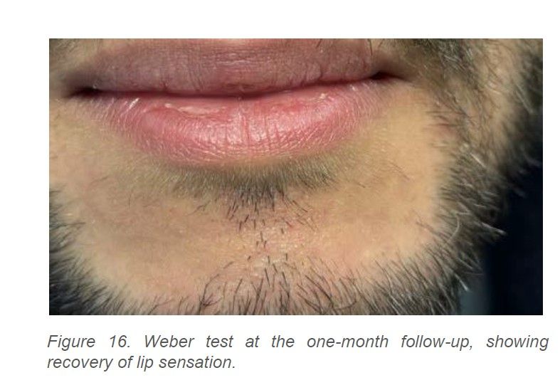
Results: After evaluating the different therapeutic options, the permanent mandibular canine was extracted and the capsule enucleated. The patient was referred to the pathologist, who confirmed the diagnosis of a dentigerous cyst. The outcome was favorable, with the only noticeable loss of lip sensation, which disappeared after one month.
Conclusions: In dentistry, it is important to detect the appearance of any lesion in the maxilla or mandible as early as possible to prevent the lesions from becoming large, thus minimizing the occurrence of complications.
Key words: Dentigerous cyst; Treatment; Complications; Nerve lesions; Pathological fractures.
Dentigerous cysts, also known as follicular, coronodental, or developmental cysts, are benign odontogenic epithelial cysts that arise from developmental abnormalities. Their formation is associated with the accumulation of fluid between the reduced enamel epithelium and the enamel surface of an unerupted tooth1.
From an epidemiological perspective, they are the second most common odontogenic cysts after radicular cysts, accounting for 14–20% of odontogenic cysts. They occur more frequently in the permanent dentition, in men aged 20–40 years, and in the mandible (70% of cases). The teeth most commonly affected are the maxillary and mandibular third molars, followed by the maxillary canines and mandibular second premolars2,3.
They are usually asymptomatic and are therefore generally diagnosed on radiographic examinations performed for unrelated reasons; however, accurate diagnosis using cone-beam computed tomography (CBCT) is required for treatment planning4.
In many cases, this diagnostic examination shows the characteristics of the radiolucent lesion, its unilocular pattern, and its central, lateral, or circumferential relationship to an impacted tooth. Depending on its growth, associated findings may include tooth displacement, root resorption of adjacent teeth, and expansion and thinning of the cortical plates5,6.
Accordingly, treatment may involve complete enucleation or more conservative techniques such as marsupialization or decompression7,8. The surgeon’s experience may be crucial, bearing in mind that mandibular lesions may be closely related to the inferior alveolar nerve or the mental nerve, either of which is generally displaced by the lesion, and that cortical weakening may even result in mandibular fracture.
The aim of this report is to present a case of a mandibular dentigerous cyst whose growth involved the mental and inferior alveolar nerves, and to analyze the treatment approach, potential complications, and their subsequent management.
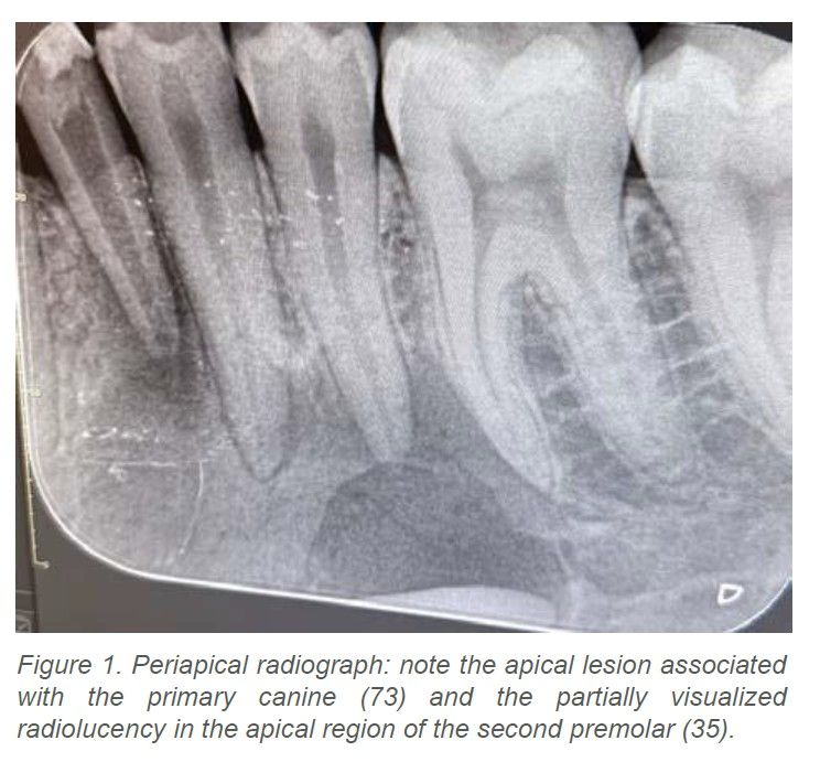
A 23-year-old man with no relevant medical history was referred by his dentist to the Oral Surgery Service at the Faculty of Dentistry, Universidad Complutense, after a periapical radiograph obtained for unrelated reasons revealed a partially visualized radiolucency in the apical region of the left mandibular premolars and primary canine (Figure 1).
During history taking, no relevant medical or surgical history was reported. The patient had no known drug allergies, reported no tobacco, alcohol, or recreational drug use, and was asymptomatic.
Extraoral examination revealed no deformity or lymphadenopathy. Intraoral examination showed that the permanent left mandibular canine was absent and the primary canine was retained. The appearance and color of the oral mucosa were normal, except for a small sinus tract overlying the apex of the primary canine. The premolars and molars showed no associated pathology, retained pulp vitality, and exhibited no mobility.
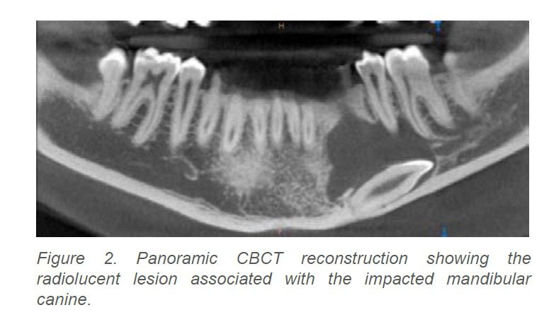
Following these findings, a CBCT examination of the entire mandible was requested. The panoramic reconstructions revealed a well-defined radiolucent lesion with an almost complete sclerotic rim, extending mesiodistally from the distal aspect of the primary canine to the apex of the first molar, reaching the inferior border of the mandible and encompassing an impacted mandibular canine (Figure 2).
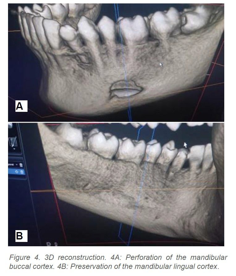
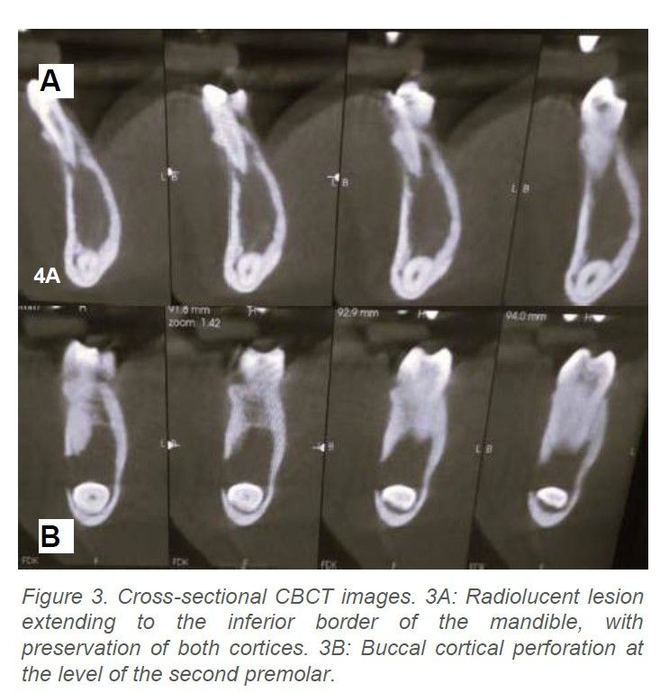
The cross-sectional CBCT images showed no cortical expansion, preservation of the lingual cortex, and slight perforation of the buccal cortex at the level of the second premolar (Figures 3A and 3B). The 3D reconstructions corroborated the findings described above (Figures 4A and 4B).
Following these findings, the patient was informed of the different treatment options and their advantages and disadvantages and elected to undergo enucleation. The patient was asked to sign an informed consent form describing the procedure and the potential intraoperative and postoperative risks. Endodontic treatment of the primary canine before surgery was considered, while the vital premolars involved in the lesion were to be monitored.
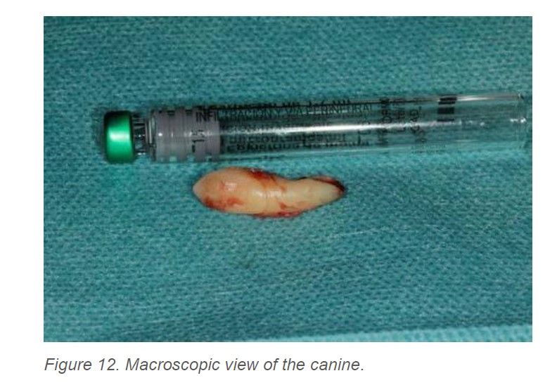
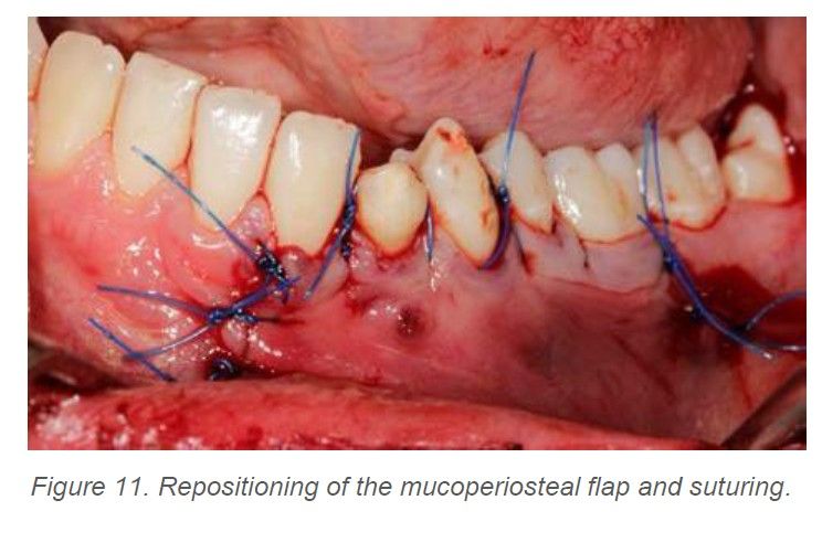
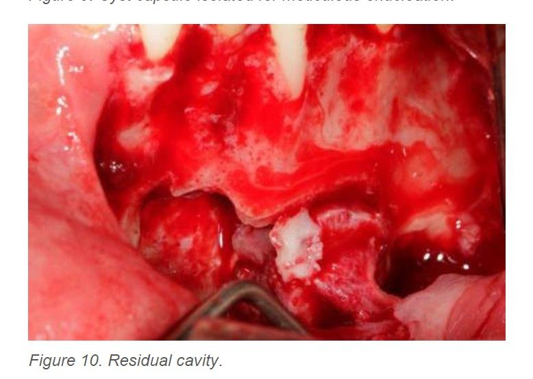
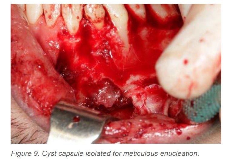
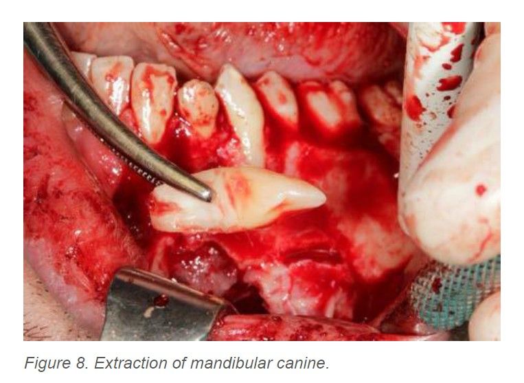
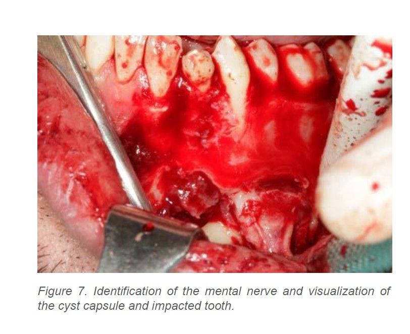
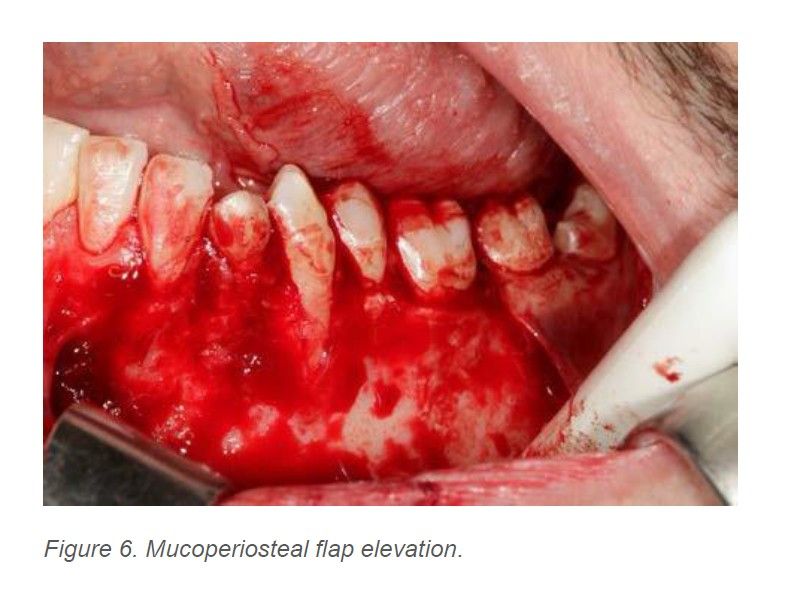
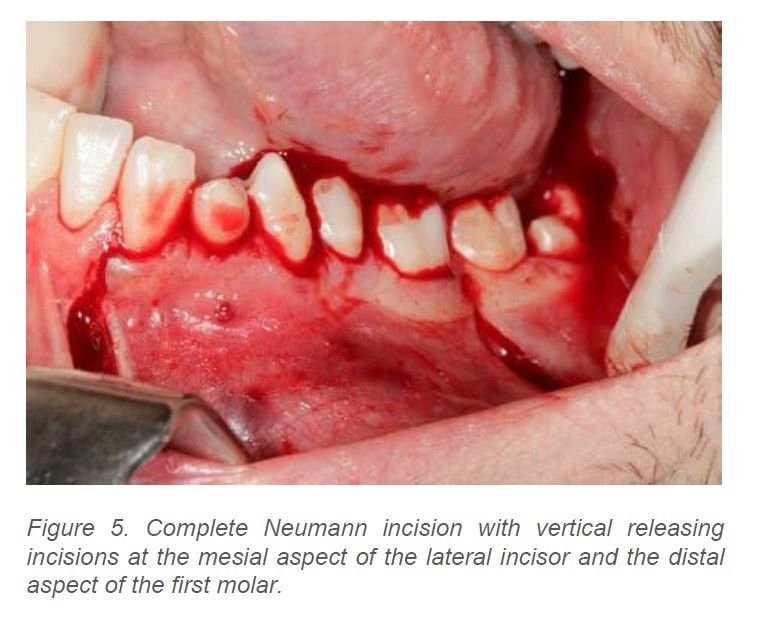
Surgical treatment was performed under sedation combined with local anesthesia administered via inferior alveolar, lingual, and mental nerve blocks. A buccal approach was used with a Neumann incision (a scalloped, marginal, full-thickness intrasulcular incision with vertical releasing incisions at the mesial aspect of the lateral incisor and the distal aspect of the first molar) followed by elevation of a mucoperiosteal flap and exposure of the mental foramen. An ostectomy was then performed to obtain the widest possible exposure of the lesion. The mandibular canine was visualized adjacent to the cyst capsule and was extracted with the mental nerve under direct visualization to facilitate complete and careful enucleation. After removal of the cyst capsule, the residual cavity was carefully curetted. Curettage was avoided along the inferior border of the mandible to prevent injury to the inferior alveolar nerve. Once the procedure had been completed, the mucoperiosteal flap was repositioned and a watertight closure was achieved using 4-0 monofilament sutures (Figures 5–12).
The specimen was placed in 10% formalin and submitted for histopathological examination. The following findings were reported:
–
Macroscopic description: Two irregular, membranous, brownish fragments with blackish areas were submitted, measuring 1 × 1.2 × 0.5 cm in aggregate. The larger fragment was sectioned, and both fragments were submitted in their entirety.
–
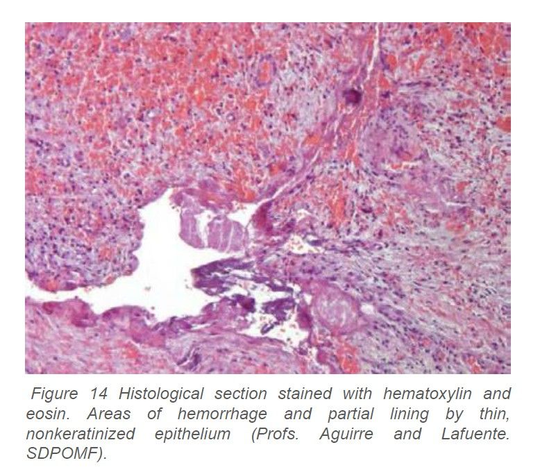
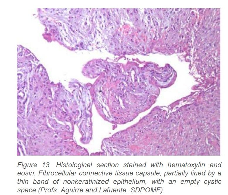
Microscopic description: Examination revealed a cystic lesion with a well-defined, thick fibrocellular connective tissue wall containing scattered vascular structures and foci of chronic lymphoplasmacytic inflammation, hemorrhage, and hemosiderin deposits. Foci of mixed inflammation with numerous foamy macrophages were also observed. In one of the fragments, a thin, nonkeratinized stratified epithelial lining was identified, with focal detachment. A peripheral shell of vital trabecular bone was observed (Figures 13 and 14). The definitive diagnosis was a dentigerous cyst with chronic inflammation.
After surgery, detailed postoperative instructions were provided and pharmacological treatment was initiated with amoxicillin 750 mg (one dose every 8 hours for 8 days), prednisone 30 mg (one dose every 24 hours for 6 days), and, as rescue analgesia, metamizole magnesium 575 mg (one dose every 8 hours as required by the patient).
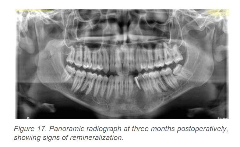
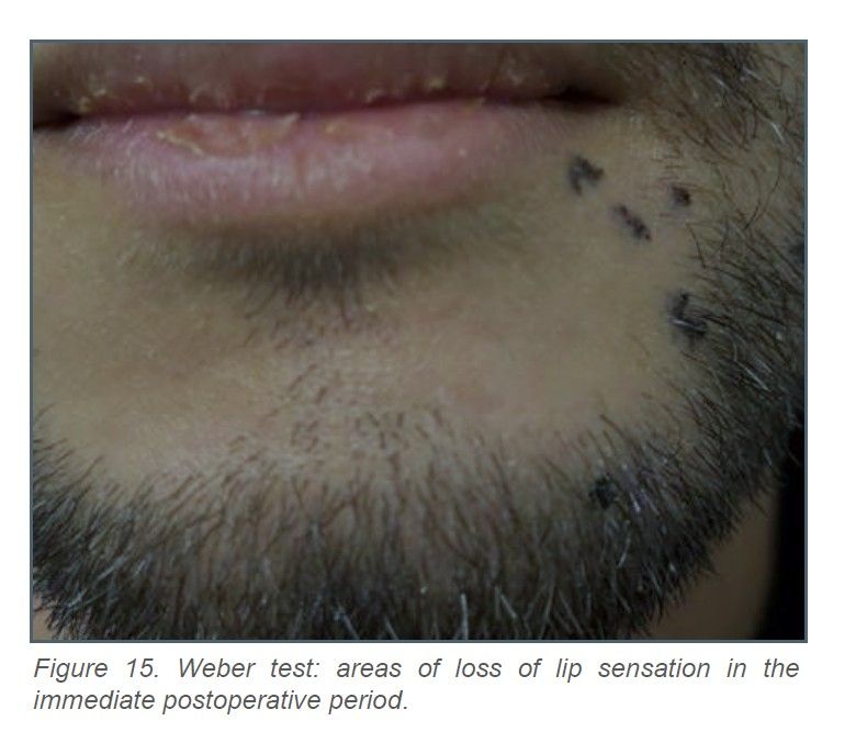
Postoperative check-ups were carried out at 2 and 7 days to review the surgical wound, and the Weber test (two-point discrimination test) was performed to assess sensation in one half of the lip; this showed a small area of sensory loss (Figure 15). The sutures were removed at 15 days, The patient remains under follow-up, with no complications to date. A radiographic follow-up examination was performed at three months and showed signs of remineralization (Figure 17).
Dentigerous cysts are benign developmental odontogenic cysts that result from the accumulation of fluid between the reduced enamel epithelium and the crown of an unerupted tooth and are associated with impacted teeth. A recent study by Sun et al.9 of 2,872 impacted teeth found a frequency of 22.55% for dentigerous cysts associated with canines, a much higher figure than the 6% observed in association with third molars.
As Karabas et al.10 point out, there is a slight male predilection, and most cases remain asymptomatic and are detected incidentally on radiographic examination, as in the present case.
Their appearance as a well-defined unilocular radiolucent lesion associated with the crown of an impacted tooth is readily detectable on panoramic radiography, although CBCT provides greater precision in characterizing the lesion and assists with the presumptive diagnosis and treatment planning11.
Lesion size appears to be a predictive factor for possible provisional diagnoses. In their study of 153 cases of unilocular radiolucent lesions, Atarbashi-Moghadam et al.12 considered that lesions larger than 2.0 cm were 5.5 times more likely than smaller lesions to be diagnosed as lesions not suggestive of dentigerous cysts. The present case meets these characteristics, which is why the patient was informed beforehand of some possible differential diagnoses, in agreement with Austin et al.13, who state that the differential diagnosis should include at least unicystic ameloblastoma and odontogenic keratocyst.
Another aspect to consider is the approach to be taken with the teeth involved in the cystic lesion. According to Zhao et al.14 and Diarra et al.15, in the presence of large cysts, it is essential to carry out an accurate assessment of pulp vitality or blood supply, both in teeth involved in the cystic lesion and in those that may lie within the surgical field.
The response of the pulp to trauma may vary, so that in some cases the pulp remains completely normal, with no adverse effects, whereas in other cases it becomes necrotic. Pulp necrosis may occur as a sequela of trauma and may promote the development of a periapical lesion due to microbial infection. Some studies, as noted by Niu et al.16, have shown that, in some cases, no pulpal infection occurred after enucleation, even in vital teeth that had not undergone root canal treatment and were affected by developmental odontogenic cystic lesions.
In case of doubt, it is recommended that root canal treatment not be performed on involved vital teeth and that follow-up be carried out over a prolonged period, making every effort to preserve their vitality17.
After this initial assessment, the size of the lesion and its relationship to surrounding anatomical structures may determine the choice of surgical treatment. The choice between enucleation, marsupialization, and decompression will often depend on the risk of intraoperative complications, as assessed by the surgeon; the surgeon’s experience will therefore be crucial.
In the present case, based on our experience, the main risk was injury to the mental and inferior alveolar nerves, given the very close relationship of the impacted tooth and the cyst capsule to these nerves.
In a review of 70 articles on displacement of the mandibular canal, Mortazavi et al.18 found dentigerous cysts to be one of the main causes.
This close relationship, among other factors, has led to the wider use of techniques such as decompression or marsupialization to prevent injury19-21. Marsupialization is increasingly recommended in very young patients with mixed dentition when the aim is to promote eruption of the involved tooth and reduce the bone damage caused by ostectomy22. In a recent systematic review of dentigerous cysts in pediatric patients, Cobo-Vázquez et al.23 found that outcomes with marsupialization or decompression were highly predictable and that these techniques facilitated eruption of the involved tooth in 83% and 100% of cases, respectively.
Despite these results, because the patient in the present case was not pediatric and the canine had been displaced toward the inferior border of the mandible, cyst enucleation and extraction of the impacted canine were chosen.
In the immediate postoperative period, as the patient had been informed before surgery, he had a small area of loss of sensation. In a study of 249 patients, Lee et al.²⁴ found that 15.26% developed inferior alveolar nerve paresthesia, showing that surgical manipulation itself almost inevitably entails some loss of sensation, which would initially be expected to be temporary.
During this period, patients may experience various neurosensory disturbances, including hypoesthesia, hyperesthesia, paresthesia, and anesthesia. These may range from minimal impairment to severe conditions and may affect the patient’s quality of life. Therefore, although there is still no standardized protocol for managing patients with paresthesia caused by nerve injury, dentists should at least be familiar with pharmacological treatments such as B vitamins and with laser therapy, as well as other approaches such as acupuncture, electrical stimulation, physical therapy, and moist heat25-27.
According to the study of refractory hypoesthesia by Hasegawa et al.28, vitamin B12 should be administered early in all cases of hypoesthesia because it may help restore axonal transport and is effective in treating neuralgia caused by traumatic nerve compression.
Likewise, laser photobiomodulation, or low-level laser therapy, has become an increasingly used adjunctive treatment.
Brito et al.29 report that this nonthermal light therapy helps modulate tissue metabolism using red or infrared light delivered by lasers or light-emitting diodes (LEDs), promoting sensory recovery after nerve injuries.
Another complication that may occur during surgical treatment is mandibular fracture. These pathological fractures are uncommon, accounting for approximately 2% of mandibular fractures, and occur most often in the mandibular angle and body30,31.
In the previously cited study of 249 patients with cystic lesions by Lee et al.²⁴, four pathological fractures occurred, representing 1.60%.
Authors such as Bazin et al.32 suggest placing fixation plates to prevent these fractures. No plate was placed in the present case because CBCT showed that, despite the large size of the lesion, the lingual cortex was completely preserved. The patient was therefore advised only to follow a soft diet for at least four weeks and avoid high-impact sports.
Finally, once the histopathological diagnosis has been confirmed, clinical and radiographic follow-up is important to confirm the absence of symptoms and remineralization at the lesion site after enucleation.
The risk of neoplastic transformation arising from odontogenic cysts is below 3%, with chronic inflammation being a key factor. In their systematic review, Sulistyani et al.33 consider that the inflammatory process can trigger genetic instability in cells through the formation of reactive oxygen species. It also induces apoptosis, cytokine production, and keratinization of the cystic epithelium and may cause alterations in DNA, proteins, and cell membranes, thereby promoting the transformation of normal cells into neoplastic cells.
It is important to detect any lesion in the maxilla or mandible as early as possible to prevent it from becoming large.
Early diagnosis, together with a precise surgical approach, will minimize risks and prevent complications such as root resorption, damage to neurovascular structures, or pathological fractures.
Follow-up is important to confirm resolution through clinical and radiographic examinations.

Sharif Ardestanian Moghadam, Alireza
Dentistry degree student. Faculty of Dentistry.
Universidad Complutense de Madrid (UCM).
Amirbagloy Darian, Melika
Dentistry degree student. Faculty of Dentistry.
Universidad Complutense de Madrid (UCM).
Martínez Rodríguez, Natalia
Part-time Lecturer in Oral Surgery. Faculty of Dentistry. Universidad Complutense de Madrid (UCM).
Miró Cuadrado, Blanca
Dentist with a Master’s degree in Endodontics. Faculty of Dentistry. Universidad Complutense de Madrid (UCM).
Martinez-Gonzalez, José María
Associate Professor of Maxillofacial Surgery. Faculty of Dentistry. Universidad Complutense de Madrid (UCM).
Correspondence:
Prof. Dr. José María Martínez-González
[email protected]