
Clinical case
Detection of carotid calcifications by panoramic dental radiography. A general health promotion action from the dental clinic
Introduction: Ischemic stroke is one of the leading causes of disability and mortality in adults. Arterial stenosis caused by carotid atheromas is a primary cause, making early identification crucial.
Aim: Although Doppler ultrasound is the most reliable diagnostic method, oral panoramic radiography can be effective preliminary detection tool.
Methods: The article presents three clinical cases where incidental detection of carotid calcifications in patients with periodontitis during routine radiographic examinations and their referral to a family physician allowed for preventive measures.
Results and conclusions: The dentists could actively contribute within the multidisciplinary medical team to the prevention of cerebrovascular accidents.
Key words: Carotid atheromas; Carotid calcifications; Carotid bifurcation; Panoramic radiographs; Stroke; Cerebral infarction; Oral health.
Cerebral infarction, or ischemic stroke, is the leading cause of disability among adults in Europe and the second leading cause of death in developed countries1,2. According to the latest estimates, one in four people worldwide will experience a stroke at some point in their lifetime3. Stroke can be classified into two main types: ischemic stroke, which accounts for 80% of cases and occurs when blood flow to part of the brain is interrupted because of a blocked blood vessel; and hemorrhagic stroke, which accounts for the remaining 20% and occurs when a blood vessel ruptures, causing bleeding in the brain and damage to the surrounding brain tissue4.
Although stroke most commonly occurs after the age of 65 years, its incidence is increasing among younger people. In recent years, a 25% increase has been recorded among individuals aged between 20 and 64 years.
According to data from the Spanish Ministry of Health, approximately 120,000 people experience a stroke in Spain each year, around 25,000 of whom die. In addition to its high morbidity and the substantial associated healthcare costs, it is important to emphasize that this disease has a profound impact on every aspect of life, affecting both those who experience it and their families5.
One of the main causes of ischemic stroke is arterial stenosis caused by the formation of atheromas in the carotid arteries. Carotid atheromas are plaques composed of lipids and inflammatory cells that may thicken, calcify and project into the vascular lumen over time, thereby altering blood flow. These plaques may also release fragments that form clots and may obstruct the intracranial arteries, triggering a stroke. Between 5% and 10% of individuals over 65 years of age have asymptomatic carotid stenosis of more than 50%; therefore, early identification of these calcifications could prevent many strokes5.
Risk factors associated with atheroma formation include a history of heart disease, physical inactivity, obesity, arterial hypertension, elevated serum cholesterol and triglyceride levels, hyperglycemia, smoking, excessive alcohol consumption and an unhealthy diet. Preventive action targeting lifestyle and modifiable risk factors could prevent up to 80–90% of cases6. However, it is important to note that in most cases, the symptoms of atherosclerosis appear at a late stage or may even be absent. The Stroke Action Plan for Europe 2018–2030 addresses the management of cerebrovascular disease from a multidisciplinary perspective, emphasizing the importance of effective coordination among the numerous professionals involved in patient care⁵.
Given that prevention is clearly the best approach, a diagnostic method capable of detecting atherosclerotic plaques before symptoms appear or blood flow is reduced would be highly valuable, as it would help prevent their progression and the development of subsequent complications.
The most widely used and reliable diagnostic method for detecting carotid atheromas is Doppler ultrasonography. This technique not only evaluates the presence of fatty plaques or clots in the carotid arteries but also determines whether blood flow is normal or obstructed, which may increase the risk of stroke.
Panoramic radiography is one of the most widely used diagnostic examinations in dental practice. It provides extensive information about the oral and facial structures, as well as the region of the upper cervical vertebrae. In this regard, Friedlander and Lande noted in 1981 that dentists have an important opportunity to contribute to the early detection of carotid calcifications in their patients, findings indicative of a high risk of stroke, simply by carefully examining the panoramic dental radiographs routinely obtained for them7.
Available studies agree that radiopaque findings consistent with carotid calcifications observed on panoramic radiographs at the level of the second to fourth cervical vertebrae are generally subsequently confirmed as carotid atheromas by Doppler ultrasonography7,12.
These calcifications, which are generally located at the carotid bifurcation, appear as radiopaque masses in the region of the panoramic radiograph corresponding to the soft tissues of the neck, approximately at the level of the C2, C3 and C4 cervical vertebrae and 1–4 cm posteroinferior to the mandibular angle13. They may vary in size and shape and may be vertically oriented and linear, nodular or heterogeneous. They may be unilateral or bilateral and single or multiple13,15.
Some studies also indicate that carotid artery calcifications are more frequent in patients with periodontitis13.
The aim of this article is to contribute to the prevention of cerebrovascular disease from the dental clinic by presenting three clinical cases in which carotid artery calcifications were incidentally detected on follow-up panoramic radiographs obtained from patients with periodontal disease undergoing maintenance therapy.
Patient 1
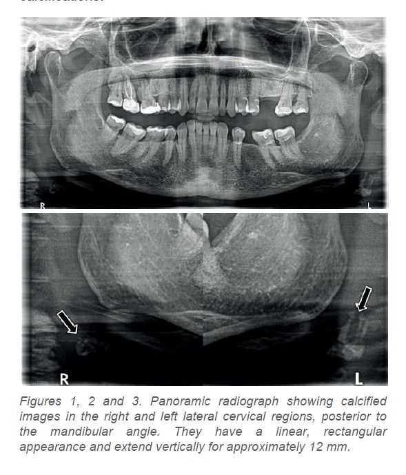
We present a 75-year-old woman with generalised stage III, grade A periodontitis. She has received periodontal treatment at our practice, where she continues to attend regular follow-up appointments. In July 2022, she attended a periodontal maintenance appointment. A follow-up panoramic radiograph was requested to assess the stability of her alveolar bone levels and rule out caries or other oral diseases. The radiograph revealed bilateral, irregularly shaped radiopacities with a linear, vertical configuration in the soft tissues of the neck, below the mandibular angle and between the third and fourth cervical vertebrae (Figures 1, 2 and 3). The presumptive radiographic diagnosis was bilateral carotid artery calcifications.
The patient’s medical history included a family history of Alzheimer’s disease. She reported that she was a non-smoker and had osteoporosis treated with denosumab administered by subcutaneous injection every 6 months. She also reported that some previous blood tests had shown slightly elevated cholesterol levels, although she was unable to provide further details or copies of the test results. She had not been prescribed any specific cholesterol-lowering treatment and occasionally consumed Danacol®.
She was referred to her primary care physician, with an explanation of the incidental finding on the panoramic radiograph, so that the physician could consider requesting further diagnostic tests to confirm the presence of carotid atheromas. The physician requested Doppler ultrasonography, which revealed several calcified atheromatous plaques at the bifurcations of both carotid arteries and at the origins of the internal carotid arteries. These calcifications were not yet causing hemodynamically significant stenosis.
Blood tests were also requested and showed elevated cholesterol levels, for which her physician prescribed statin therapy.
In this patient, the diagnosis made by her physician was early and preceded the onset of symptoms, owing to the detection of the calcifications on a panoramic radiograph obtained during periodontal maintenance.
Patient 2
We present a 77-year-old woman who smokes 10 cigarettes per day and has generalised stage IV, grade C periodontitis. She has received periodontal treatment at our practice, where she continues to attend regular follow-up appointments. At her most recent maintenance appointment in 2024, when asked whether there had been any changes in her medical history, she reported that she had recently been diagnosed with calcifications in the left carotid artery after lower blood pressure had been observed in her left arm than in her right arm. In view of this finding, her family physician requested Doppler ultrasonography, which revealed the calcified atheromatous plaques.
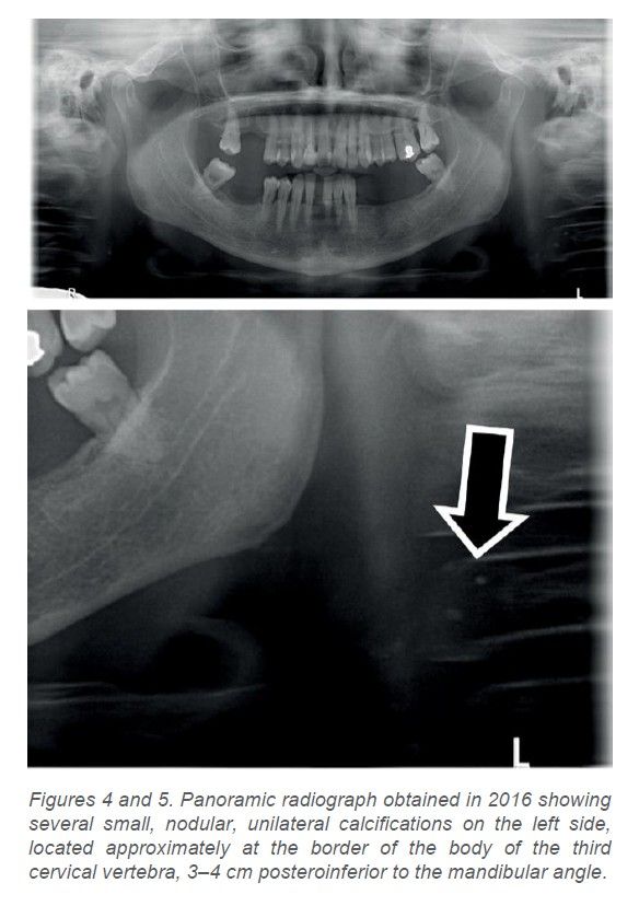
Following the patient’s explanation, her most recent panoramic radiograph, taken in 2016, was reviewed. Several small, round, nodular calcifications were already visible in the region of the second to fourth cervical vertebrae, although they had gone unnoticed at the time (Figures 4 and 5).
In this patient, the carotid atheromas were diagnosed after the onset of symptoms, although fortunately before any occlusive cardiovascular or cerebrovascular event had occurred. This case highlights the importance of identifying such images on panoramic radiographs, as this may allow early diagnosis and consequently help prevent associated complications.
Patient 3
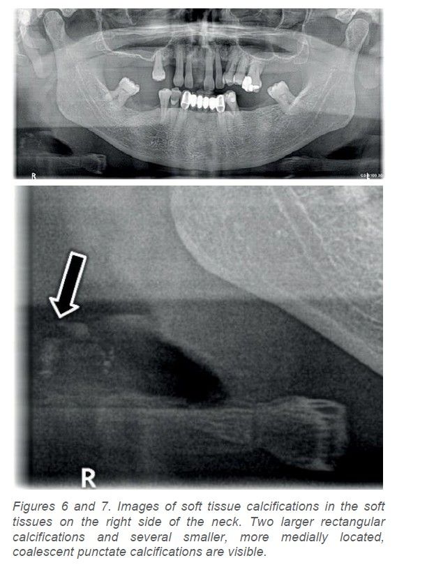
We present a 64-year-old man with generalised stage IV, grade B periodontitis. He has received periodontal treatment and has been undergoing periodontal maintenance for 20 years. His relevant medical history included hepatitis C, for which he was not currently receiving treatment; ankylosing spondylitis, for which he received no regular treatment other than non-steroidal anti-inflammatory drugs during episodes of joint pain; and controlled hypertension treated with ramipril. At a periodontal maintenance appointment, a follow-up panoramic radiograph was obtained. It showed two rectangular calcified images measuring approximately 5 mm in the soft tissues on the right side of the neck, together with smaller punctate images located more medially, approximately 3–4 cm from the mandibular angle (Figures 6 and 7). Because carotid calcifications were suspected, the patient was referred to his primary care physician to assess the need for specific diagnostic testing. A vascular surgeon requested Doppler ultrasonography, which confirmed a predominantly calcified atheromatous plaque at the right carotid bifurcation. The plaque measured approximately 5 × 1.5 mm and was not causing hemodynamically significant stenosis. Blood tests were also requested and revealed no significant abnormalities.
In this patient, as in Patient 1, carotid atheromas were diagnosed before the onset of symptoms and before the development of carotid stenosis, which is essential for preventing cerebrovascular events.
All three patients were informed through the informed-consent process that their data could be used for research purposes.
The use of panoramic radiographs as a diagnostic tool in dentistry is common because of the large amount of information they provide, their ease of acquisition and their low cost. The scientific literature recognizes their effectiveness in visualizing cervical calcifications8,16.
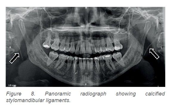
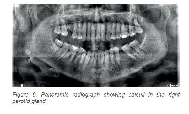
When a calcification is suspected during the assessment of a panoramic radiograph, it is essential to establish a differential diagnosis with other radiopacities, whether anatomical or pathological. First, normal anatomical structures must be identified, including the hyoid bone, epiglottis, stylomandibular ligaments (Figure 8), stylohyoid ligaments and triticeal cartilage, which may sometimes appear calcified. They must then be differentiated from pathological processes, including other types of calcification that may be caused by carotid artery atheromas or by other conditions such as salivary gland calculi (Figure 9), phleboliths, tonsilloliths or even calcified cervical lymph nodes16.
In 1998, Friedlander and Friedlander identified the sites most frequently affected by atheromatous lesions as the apex of the common carotid artery bifurcation and the posterior wall of the proximal internal carotid artery⁸. Radiographically, these areas correspond to two projections: one bounded anteriorly by the mandibular ramus and mandibular angle and posteriorly by the bodies of the 2nd, 3rd and 4th cervical vertebrae, and another located at the inferior border of the body of the third cervical vertebra, 1.5–4 cm inferior and posterior to the mandibular angle11,17.
When small, they have a circular radiographic appearance. Large atheromas, however, have a linear or thin rectangular shape. In Patient 1, the calcifications were bilateral, large, rectangular and irregular, located 1–2 cm posteroinferior to the mandibular angle and superimposed over the prevertebral soft tissues. In Patient 2, however, the calcifications were unilateral, smaller, circular and multiple, superimposed over the prevertebral soft tissues and adjacent to the hyoid bone.
In Patients 1 and 3, the calcifications were located at the carotid bifurcation, the most frequent location according to Friedlander and Friedlander⁸. In Patients 2 and 3, the carotid atheromas were unilateral: on the left in Patient 2 and on the right in Patient 3. These findings corresponded to the unilateral calcifications observed on the panoramic radiographs obtained solely for periodontal follow-up.
Several publications have analyzed calcifications detected on panoramic radiographs in the region of the soft tissues of the neck superimposed over the carotid arteries and subsequently confirmed as carotid artery calcifications by Doppler ultrasonography. In 2005, Friedlander et al. were among the first authors to report this finding after analyzing 1,548 consecutive panoramic radiographs from neurologically asymptomatic patients over 50 years of age. They detected atheromas in 65 of these patients, which were subsequently confirmed by Doppler ultrasonography9.
A relevant cross-sectional study because of its sample size was conducted in 2022 by Janiszewska-Olszowska et al., who analyzed 4,000 panoramic radiographs from patients aged between 45 and 87 years, with a mean age of 62 years. The study found that the mean age at which calcifications appeared ranged from 61 to 65 years. Calcifications were identified in 21.68% of cases, comprising 12.97% in women and 8.4% in men14.
Calcifications may occur unilaterally or bilaterally. In 2019, Garoff et al.18 concluded that bilateral calcifications with a vessel-outlining morphology, as in Patient 1, are independent risk markers for future cardiovascular events.
Similarly, a 2023 Swedish study by Bladh et al., which included 414 participants, concluded that, irrespective of their morphology, all carotid artery calcifications detected on panoramic radiographs were associated with a greater degree of cardiovascular disease findings on Doppler ultrasonography than in patients without calcifications on panoramic radiographs12.
Some studies, such as that conducted by Madden et al. in 2007 in 54 patients, have found that panoramic radiographs may have relatively low sensitivity for detecting carotid artery calcifications compared with ultrasonography19. Nevertheless, when vascular examination by Doppler ultrasonography is performed following the incidental discovery of a radiopacity in the carotid region on a panoramic radiograph12,16, the sensitivity of panoramic radiography for this indication improves significantly.
The aim of this article is not to promote the systematic use of panoramic radiography as a routine screening method for detecting carotid artery calcifications, but rather to emphasize the important contribution dentists can make to their early detection, potentially helping to save a life through a panoramic radiograph obtained for dental reasons17. Accordingly, all structures should be examined carefully when reviewing routine panoramic radiographs, without limiting the examination to the oral cavity, particularly in patients with periodontitis13,14 or in older patients with risk factors such as smoking, arterial hypertension, diabetes mellitus, obesity or hyperlipidemia10,20.
Once a calcification has been detected on a panoramic radiograph, the patient must be referred to their primary care physician or cardiologist so that the appropriate tests, such as Doppler ultrasonography, can be performed to confirm or exclude the risk of cerebrovascular disease. When a panoramic radiograph is obtained, it is primarily examined for dental and bone pathology, but it is also essential to assess the other anatomical structures visible on the image, both in the vertebral region and in the soft tissues of the neck. In reality, however, many dentists are not fully aware of these areas when interpreting panoramic radiographs. Nevertheless, with some training and careful examination, calcifications can be detected in approximately 3–15% of patients⁹. These calcifications could indicate an increased risk of stroke12,15,18. Furthermore, as artificial intelligence models continue to be developed, the accuracy with which these calcifications are detected on panoramic radiographs will increase in the future21. It is therefore essential to reinforce the importance of examining the oropharynx and identifying possible lesions in both undergraduate and postgraduate education22.
The management of carotid stenosis is a multidisciplinary process involving the primary care physician, radiologist, cardiologist and vascular surgeon. The dentist can, however, play a key role in this process by detecting calcifications early on panoramic radiographs, thereby contributing to early diagnosis by the physician and helping prevent their possible progression to stroke. The oral healthcare team should also help reduce risk factors shared by oral and cerebrovascular diseases, including smoking, a diet high in sugars and saturated fats, alcohol consumption, obesity and physical inactivity, within the multidisciplinary preventive framework recommended by the Spanish Ministry of Health.

Serrano Sánchez-Rey, Mª Cristina
Master’s degree in Periodontology from Universidad Complutense de Madrid (UCM). Member of the SEPA–SED Working Group on Diabetes and Periodontitis.
Private practice in periodontology and implant dentistry in Ciudad Real.
Rubio Ramírez, Lourdes
University Specialist in Oral Medicine, Universidad Complutense de Madrid (UCM). Contributing lecturer on the UCM Specialist in Oral Medicine programme. Private practice in periodontology in Ciudad Real.
Casas Hernández, Agustín
Master’s degree in Periodontology from Universidad Complutense de Madrid (UCM). Private practice in periodontology and implant dentistry in Alcorcón.
Carasol Campillo, Miguel
Physician and specialist in stomatology. PhD in Dentistry. Coordinator of the Alliance for Periodontal and General Health at Fundación SEPA.
Alonso Álvarez, Bettina
Associate Professor. Department of Clinical Dental Specialties. Faculty of Dentistry, Universidad Complutense de Madrid (UCM).
Correspondence:
Cristina Serrano Sánchez-Rey
C/ Ramón y Cajal, 3, 2º A
13001 Ciudad Real
Tfno 926256459