
Clinical report
Microinvasive approach to the management of white spot lesions in a case of amelogenesis imperfecta
Introduction: White spot lesions are opacities in the dental enamel as a consequence of a defect in its mineralization. This hypomineralization can be an initial sign of a carious lesion, or due to a defect in the enamel as a consequence of an alteration during its formation. The etiology and extent of the lesions determine the therapeutic approach. The aim of the present case report is to describe the microinvasive approach of a case of amelogenesis imperfecta in a mild form of presentation.
Materials and Methods: A clinical case of a 33-year-old non-smoker female patient with no previous registered medical records (ASA I), with the presence of white spot lesions as a clinical manifestation of amelogenesis imperfecta hypomature type, subtype IIC, that were treated by a microinvasive approach combining bleaching which improves the optical properties of the lesion as it has a refractive index similar to that of healthy enamel.
Results: Dental bleaching is a non-invasive procedure, which in this case partially masked the white spot lesions and improved the substrate for subsequent resin infiltration. Infiltration is a microinvasive, fast and safe procedure,which allows good esthetic results achieving complete masking of most lesions.
Conclusions: Resin infiltration combined with bleaching is a conservative procedure that respects tooth structure and provides satisfactory esthetic results in the treatment of white spot lesions caused by amelogenesis imperfecta.
KEY WORDS: White spot lesions; Amelogenesis imperfecta; Resin infiltration; Tooth bleaching; Microinvasive treatment.
White spot lesions (WSLs) are defined as non-cavitated whitish opacities on the surface of dental enamel resulting from enamel hypomineralization or hypomaturation1,2. The presence of these lesions, particularly on anterior teeth, may cause significant aesthetic alterations and may even affect patients’ self-esteem. WSLs have multiple etiological factors and may develop during either the pre-eruptive or post-eruptive phase. The former group includes dental fluorosis, traumatic hypomineralization, molar-incisor hypomineralization (MIH) and amelogenesis imperfecta, whereas the latter includes initial caries lesions. Regardless of their etiology, all WSLs involve a reduction in subsurface enamel mineral content, resulting in changes in its chemical composition and consequent alterations in its optical properties3.
The clinical features of WSLs include loss of enamel translucency, an opaque white appearance, particularly when the tooth is dehydrated, and a porous, rough surface. This mineralization defect and a possible reduction in the density of the surface enamel layer may be detectable radiographically or by transillumination2.
The characteristic opacity of WSLs is caused by increased microporosity between the hydroxyapatite crystals, with the resulting spaces being occupied by organic fluids that alter the refractive index (RI) of enamel.
Healthy enamel has an RI of 1.62, whereas organic fluids, which consist mainly of water, have an RI of 1.33. This difference in RI within the same tissue alters the perception of light and is the physical phenomenon responsible for the optical changes observed in enamel affected by WSLs. When the enamel is dehydrated, these fluids are replaced by air, which has an RI of approximately1. As this value differs more markedly from that of healthy enamel, the WSL becomes more evident in the dehydrated tooth and appears whiter and more opaque4,5. It should be noted that the surface layer of some lesions may be hypermineralized, particularly in long-standing lesions or lesions that have been treated with remineralizing agents, which may influence the treatment approach.
Determining the etiology, size and depth of WSLs is essential before selecting a treatment option, as treatment outcomes vary according to the enamel substructure1 and the patient’s aesthetic expectations. A thorough visual examination allows the main characteristics of WSLs to be assessed; however, supplementary diagnostic methods such as photography and, particularly, transillumination enable their shape, size, color, location, depth and extent to be evaluated more accurately⁵.
Pre-eruptive WSLs result from disturbances during amelogenesis, either during the secretory phase or the maturation phase, producing enamel of normal volume but with a lower degree of mineralization and, therefore, qualitative defects. This article focuses on one of the conditions included in this group: amelogenesis imperfecta (AI)6.
Amelogenesis imperfecta (AI) is a disorder of enamel formation and mineralization characterized by hypoplasia and/or hypomineralization. It is genetic in origin and may be inherited in an autosomal dominant, autosomal recessive, or X-linked dominant pattern; sporadic cases also occur. It affects the structure and clinical appearance of the enamel of all or nearly all teeth in a similar manner. One of the most widely used classifications is that proposed by Witkop in 1988, which takes into account the enamel phenotype and mode of inheritance (Table)7. As this classification shows, AI is a complex disorder that may have different clinical expressions or forms. Because of its genetic origin, it affects both the primary and permanent dentitions, irrespective of the chronology of tooth eruption6,8,9.
This developmental enamel defect results from pathogenic variants in, or altered expression of, genes encoding proteins involved in amelogenesis, including enamelin (ENAM), amelogenin (AMELX), enamelysin (MMP20), kallikrein-related peptidase 4 (KLK4), FAM83H, WDR72, FAM20A, SLC24A4, DLX3, AMBN, LAMB3, ITGB6, and C4orf26. The possible roles of ALB and TUFT1 in amelogenesis are also under investigation.
These genetic alterations disrupt normal ameloblast function, resulting either in enamel of reduced thickness in the hypoplastic forms of AI or in enamel of normal thickness but with defective mineralization and maturation in the hypocalcified or hypomaturation forms10,11. The clinical presentation of AI depends on the gene involved and the stage of amelogenesis at which the defect is expressed. It may range from superficial changes in color to complete enamel aplasia12. and may be accompanied by aesthetic impairment and hypersensitivity12,13. Although AI is primarily an enamel-specific disorder, it is frequently associated with other abnormalities, including pulp calcifications, delayed tooth eruption, congenitally missing teeth, root resorption, open bite, negative overjet, and discrepancies in the vertical relationship of the jaws9,14,15.
This article focuses on type II hypomaturation AI, which presents a less severe qualitative enamel defect because the disturbance occurs during the maturation stage of amelogenesis. It is characterized by reduced enamel mineral content. The enamel may be less translucent, opaque, and pigmented; it is harder than in the hypocalcified forms and with a normal thickness. The enamel surface is usually smooth, although it may appear mottled or show vertical bands of alternating normal and highly permeable hypomature enamel12,15.
Enamel discoloration may be white, yellowish, grayish, or brownish. The mildest form of this type of AI often goes undiagnosed, and patients generally seek treatment for aesthetic reasons15. This type of AI includes a clinical variant known as “snow-capped enamel” or “snow-capped teeth,” in which an area of opaque whitish enamel is present in the occlusal or incisal third of the teeth. Because of the clinical appearance of the teeth, it may sometimes be necessary to differentiate this condition from dental fluorosis14.
The principal treatment options for WSLs of pre-eruptive origin include tooth bleaching, which is generally the first step in almost all cases, followed by or combined with enamel microabrasion, resin infiltration, or, where necessary, direct composite restorations.
Tooth bleaching does not treat the lesion itself; its main purpose is to mask it by reducing the contrast between the lesion and the surrounding sound enamel. Its principal advantage is that it is a non-invasive procedure that also provides a more favorable aesthetic substrate for subsequent procedures16. Its disadvantage is that it does not usually produce satisfactory results when used alone.
Enamel microabrasion is particularly effective for superficial WSLs. This procedure combines abrasive agents, such as pumice or silica, with an acid, such as 37% phosphoric acid or 6% hydrochloric acid. One disadvantage is that it removes a greater thickness of enamel than resin infiltration; it may also cause sensitivity in some cases, reduce enamel thickness and even initiate the restorative cycle5,17. It is important to note that all these procedures may be combined to obtain better aesthetic results while remaining as conservative as possible.
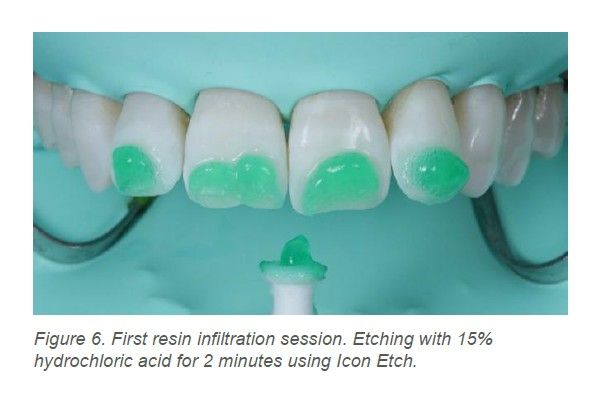
Resin infiltration is a microinvasive procedure initially used to treat non-cavitated proximal caries lesions, and its use has expanded in recent years to the aesthetic treatment of WSLs. The procedure is based on etching the enamel surface with 15% hydrochloric acid for 2 minutes, thereby increasing the porosity of the surface enamel, followed by dehydration with 99% ethanol and, finally, infiltration with a low-viscosity resin¹⁸, which penetrates the body of the lesion by capillary action and fills the microporosities3,4,18. It is a low-molecular-weight resin, specifically triethylene glycol dimethacrylate (TEGDMA), which improves the optical properties of the lesion because it has an RI of 1.52, similar to that of sound enamel (1.62), thereby masking the lesion. Its effectiveness in improving the aesthetic appearance of WSLs has been demonstrated³-⁵. The advantages of resin infiltration include its microinvasive nature, as only a minimal layer of enamel, 34 μm per application, is removed19,20; it does not cause sensitivity and provides better aesthetic results than other procedures. However, resin infiltration does not remineralize the enamel, and satisfactory results are not always achieved in deep lesions. It must therefore be combined with other procedures, such as microabrasion or even composite restorations15,16.
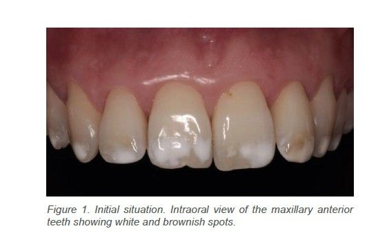
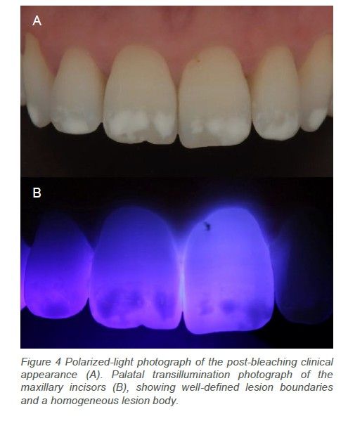
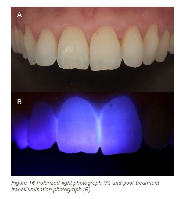
A patient was referred to the Master in Lifelong Training in Aesthetic Restorative Dentistry and Endodontics at Rey Juan Carlos University for aesthetic treatment of her WSLs. She was a 33-year-old woman with no medical conditions recorded in her medical history. She reported that she had been born in Orense and raised in Málaga and Madrid, Spain. Clinical examination revealed generalized WSLs in the incisal third of the maxillary anterior teeth and isolated lesions in the posterior regions. The lesions showed a homogeneous distribution and color pattern (Figure 1). A photographic record was also obtained using a Nikon D7200 camera and an AF-S Micro-Nikkor 105 mm lens. This was supplemented with photographs taken using a polarizing filter (Polar_eyes; Nikon R1C1) and with transillumination, for which the tip of a Mini LED light-curing unit (Acteon Satelec, France) was positioned palatally. These non-invasive adjunctive examinations made it possible to determine the extent and depth of the WSLs.
Most lesions were superficial and well demarcated, with a homogeneous lesion body (type I), according to the transillumination-based classification of Marouane and Manton (2021)²¹.
Because WSLs were present on nearly all the teeth and the information obtained during history-taking did not indicate another cause, a diagnosis of type II AI, hypomaturation subtype IIC (“snow-capped enamel” or “snow-capped teeth”), was made13,14. Because as it is a genetically determined disorder, genetic testing is required to confirm the diagnosis and determine the inheritance pattern; however, the patient did not consider it necessary to undergo this test.
These superficial, well-demarcated lesions are considered to respond better to resin-infiltration treatment than deep lesions with heterogeneous lesion bodies, which may require modifications to the infiltration protocol or more invasive restorative treatments²¹.
A combined treatment was therefore proposed, beginning with tooth bleaching followed by resin infiltration of the WSLs, as this was the most conservative option15,16.
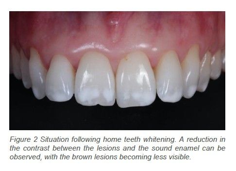
After basic periodontal treatment had been completed, at-home bleaching was initiated using 16% carbamide peroxide (Pola Night, SDI, Australia), applied overnight in custom-made trays fabricated from thermoplastic sheets. The initial shade, 3M1, was recorded using the VITA Toothguide 3D-MASTER shade guide (VITA Zahnfabrik, Bad Säckingen, Germany). The aim of bleaching was to reduce the contrast between the lesions and the sound enamel. During the final week of treatment, the patient was instructed to apply the bleaching agent every other day because she developed mild tooth sensitivity. After 4 weeks of application, a final shade of 1M1 was obtained(VITA Toothguide 3D-MASTER shade guide), and bleaching was discontinued because the aesthetic objective was considered to have been achieved (Figure 2).
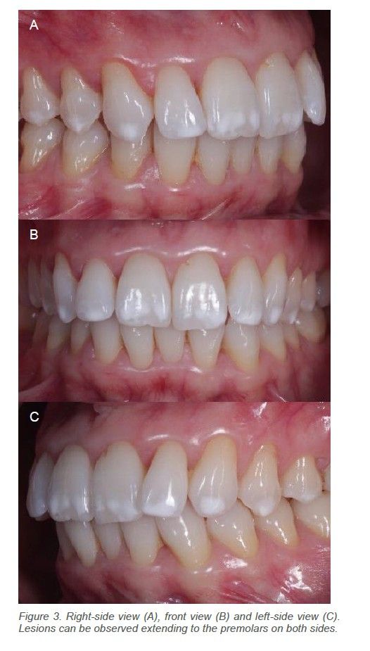
Despite the marked tooth-whitening effect, the WSLs were not completely masked (Figure 3); resin infiltration was therefore performed. Before resin infiltration, photographs were taken using a polarizing filter and transillumination to confirm the extent and homogeneity of the lesions (Figure 4).
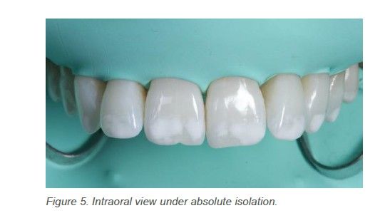
The Icon Vestibular resin infiltration kit (DMG, Hamburg, Germany) was used in accordance with the manufacturer’s instructions, with slight modifications to the protocol. Absolute isolation was achieved from teeth 15 to 25 using a Nic Tone rubber dam (MDC Dental, Mexico) and No. 00 clamps (Figure 5).
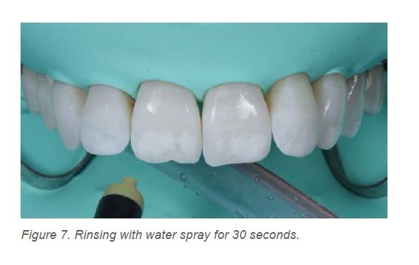
The WSLs were etched with Icon Etch, a 15% hydrochloric acid, for 2 minutes (Figures 6 and 7). The treated surfaces were then rinsed with water spray and dried. A 99% ethanol solution (Icon Dry) was then applied. Three seconds after its application, transillumination was used to determine whether the lesions disappeared, in what is known as the ethanol test19,21.
As the lesions did not disappear, the etching procedure was repeated four times. The number of applications was therefore increased beyond that recommended by the manufacturer in order to achieve deeper etching of the lesions and improve the degree of infiltration22-24. The hydrochloric acid was also applied actively to eliminate CO₂ bubbles, improve lesion conditioning²⁴ and achieve deeper removal of the superficial hypermineralized layer.
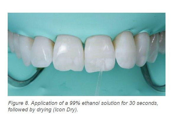
Icon Dry was then applied for 30 seconds (Figure 8), followed by drying with moisture-free air for 30 seconds. The principal function of ethanol is to dehydrate the microporosities of the WSLs and facilitate resin infiltration. It is recommended that the process be repeated at least once to remove as much water as possible. However, as noted above, it may also help predict whether the lesion can be masked by infiltration: if the lesion disappears within the first 3 seconds after being moistened with ethanol20,21,23,24.
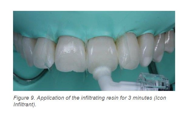
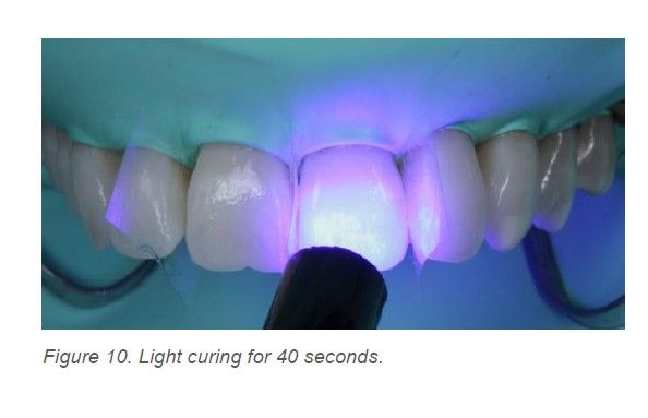
The low-viscosity infiltrating resin, Icon Infiltrant, was then applied to the white spot lesions and left in place for 3 minutes, after which it was light-cured for 40 seconds using a Mini LED light-curing unit (Acteon Satelec) (Figures 9 and 10). The procedure was repeated, allowing the infiltrant to remain in place for 1 minute, after which it was light-cured again for a further 40 seconds25.
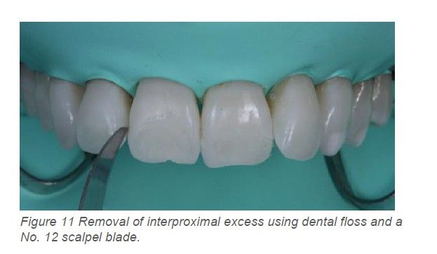
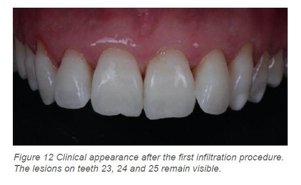
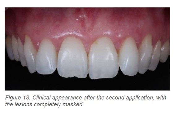
Once infiltration had been completed, the interproximal excess was carefully removed using dental floss and a No. 12 scalpel blade (Swann-Morton) (Figure 11), and the isolation was removed. The facial surfaces were polished using fine- and superfine-grit Sof-Lex discs (3M ESPE, USA), DIACOMP PLUS TWIST polishers (EVE, Germany), and Enamelize aluminum oxide polishing paste (Cosmedent,USA). At a follow-up appointment 7 days later, the lesions on teeth 23, 24 and 25 had not completely disappeared (Figure 12). A further application of Icon Vestibular was therefore performed on these teeth after roughening the lesion surfaces with a medium-grit Sof-Lex disc. The entire procedure described above was repeated, with favorable aesthetic results (Figure 13).
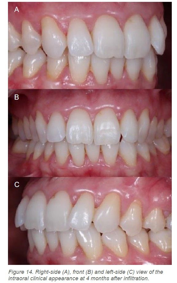
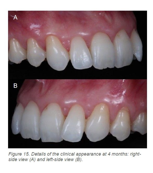
After 4 months, the results were reviewed (Figures 14 and 15), and polarized-light and transillumination photographs were taken again (Figure 16)
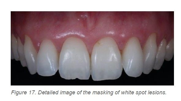
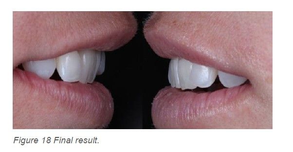
AI is a genetically determined disorder of enamel formation that, in its mild forms, may present solely as WSLs. The treatment of aesthetic alterations represents a challenge for the clinician, and the treatment approach is selected according to lesion severity, the patient’s age and the patient’s aesthetic expectations12,15,16. This case demonstrates the good aesthetic results achieved by combining tooth bleaching with low-viscosity resin infiltration (Figures 17 and 18). These microinvasive procedures should therefore be considered before conventional procedures when treating these clinical presentations of AI.
Tooth bleaching is a non-invasive procedure that, in this case, partially masked the WSLs by reducing the differences between sound and affected enamel and improved the substrate for subsequent resin infiltration16. Therefore, even when tooth bleaching does not achieve the desired result, it may improve the final outcome when combined with other procedures, as it removes other types of stains, such as the brown stains present in this case, which is necessary before the infiltration procedure26.
With regard to resin infiltration, the greatest challenge is adequately conditioning the hypermineralized surface enamel layer, because if this layer remains, the resin cannot infiltrate or mask the subsurface lesion. The use of 15% hydrochloric acid has been shown to be the best option for conditioning the surface enamel layer, removing approximately 34 μm of tooth structure with each application performed according to the manufacturer’s recommended protocol. In this case, the conditioning protocol was modified because, when the ethanol test and transillumination were performed during the first 3 seconds after application of Icon Dry, some lesions showed a negative ethanol-test pattern. It was therefore necessary to repeat the Icon Etch application procedure, as indicated by Marouane and Manton²¹. Arnold et al. (2015)22 had previously found that repeating the conditioning procedure increases the etching depth. Other authors have reported that the number of hydrochloric acid applications may be increased, provided that no more than seven applications are performed22-25. Although the number of applications recommended by the manufacturer was exceeded, resin infiltration would still be more conservative than microabrasion, which may remove up to 360 μm of enamel22, or the use of a bur.
The low-viscosity TEGDMA resin fills the microporosities of the lesion by capillary action, preventing them from being occupied by oral fluids or air. Its ability to penetrate the lesion is determined by the previous conditioning of the enamel and the resin infiltration time. Although a 3-minute application is recommended, the infiltration time may be extended to up to 15 minutes to achieve a greater degree of infiltration25,27,28. However, in our case, the application time was not extended because the ethonol test was positive, which led us to expect a good response to infiltration²¹.
The advantages of this resin infiltration procedure are that it is microinvasive, preserves tooth structure, is safe and rapid, provides good aesthetic results in a single visit, and is highly acceptable to patients29-32. One disadvantage is that it does not achieve complete masking in deep lesions and that, despite producing a good aesthetic result, its application does not remineralize the lesion but only masks it28,33.
Finally, although the color stability of infiltrated WSLs has been questioned, published clinical studies agree that the aesthetic results are maintained34, even 6 years after infiltration35.
Resin infiltration, preceded by tooth bleaching, is an effective and conservative alternative for treating WSLs caused by a mild form of AI. This therapeutic combination not only provides satisfactory aesthetic results but also preserves tooth structure. The use of ethanol and simultaneous observation of its effect using transillumination are valuable diagnostic tools that can predict the success of resin infiltration, thereby optimizing the clinical management of these lesions.


Angamarca Alvarado, Edy
Student on the Master in Lifelong Training in Aesthetic Restorative Dentistry and Endodontics. URJC
Camacho Terciado, Silvia
Student on the Master in Lifelong Training in Aesthetic Restorative Dentistry and Endodontics. URJC.
Olea Vielba, M
Lecturer on the Master in Lifelong Training in Aesthetic Restorative Dentistry and Endodontics. URJC.
Ceballos García, Laura
Director of the Master in Lifelong Training in Aesthetic Restorative Dentistry and Endodontics. URJC.
Correspondence:
Edy Angamarca Alvarado.
Calle Golfo de Salónica 8, 9C.
28033, Madrid. [email protected] [email protected]