
ORIGINAL ARTICLE
Implants 4.5mm long, used in bone atrophy at height (residual bone ≤ 4.5mm). Retrospective study
Introduction: Vertical atrophy in the posterior maxilla represents a frequent challenge in dental implant rehabilitation. Transcrestal sinus floor elevation has gained relevance in these cases. When 5.5 mm implants are insufficient due to limited bone availability, 4.5 mm extra-short implants have emerged as a viable alternative. This study presents a retrospective series of cases treated with this approach.
Methods: Patients who received 4.5 mm-long implants in residual ridges with ≤4,5 mm of bone height were retrospectively analyzed. A transcrestal sinus lift was performed using autologous bone chips obtained from the drilling process and PRGF-Endoret as grafting material. The primary variable was implant survival; secondary variables included crestal bone stability after prosthetic loading. Qualitative variables were analyzed using frequency analysis; quantitative data were described with mean ± standard deviation. Implant survival was estimated using the Kaplan-Meier method.
Results: Sixteen patients with a total of 18 implants met the inclusion criteria. The mean residual bone height at implant sites was 3.96 mm (±0.38). Sinus lifts ranged from 0.2 to 1.3 mm. All implants were 4.5 mm in length. The mean follow-up was 36.3 months (±2.71). No implant failures were recorded, resulting in a cumulative survival rate of 100%. Mean marginal bone loss at the end of follow-up was 0.35 mm mesially and 0.11 mm distally.
Conclusions: 4.5 mm implants combined with transcrestal sinus floor elevation and PRGF-Endoret appear to be a predictable therapeutic option for the rehabilitation of severely resorbed posterior maxillae.
Key words: Bone atrophy, transcrestal sinus lift, extrashort dental implant.
Transcrestal sinus floor elevation combined with extra-short implants has become a widely used approach for rehabilitating the posterior maxilla in cases of moderate-to-severe horizontal bone atrophy¹˒². Different protocols are used for the surgical approach, implant site preparation and implant placement3-6. The choice of implant type depends directly on residual bone height. Where residual bone height permits, short implants (6.5–7.5 mm) or extra-short implants (5.5–6.5 mm) can be used⁷˒⁸. However, when only 3–5 mm of bone is available, even shorter implants are required to facilitate a minimally invasive crestal approach. In situations involving less bone volume and shorter implants, achieving primary stability becomes more difficult, although it is essential for treatment success. Our research group is among those that have shown that transalveolar sinus floor elevation can be performed with short and extra-short implants even when residual ridge height is less than 5 mm, provided that the implant is adequately stabilised in three dimensions around the perimeter of the alveolar crest. To achieve this, the drilling sequence must be adapted to the characteristics of the residual bone, and implants designed to provide three-dimensional anchorage (buccopalatal and mesiodistal) must be used to achieve optimal fixation within the alveolar crest. This approach is essential to ensure stability even in challenging situations, such as those requiring an implant less than 5.5 mm long. The need to treat such cases led to the development of 4.5-mm-long implants. Few studies have evaluated implant survival and crestal bone loss in implants less than 5 mm long. Nevertheless, the available studies agree that these implants do not have higher failure rates or greater bone resorption than standard-length implants placed in conjunction with regenerative procedures in cases of severe bone atrophy or with procedures such as transcrestal sinus floor elevation. The present study reports a retrospective case series in which 4.5-mm-long implants were placed in the posterior maxilla at sites with a residual bone height of ≤5 mm, using particulate autologous bone combined with PRGF-Endoret as the graft material.
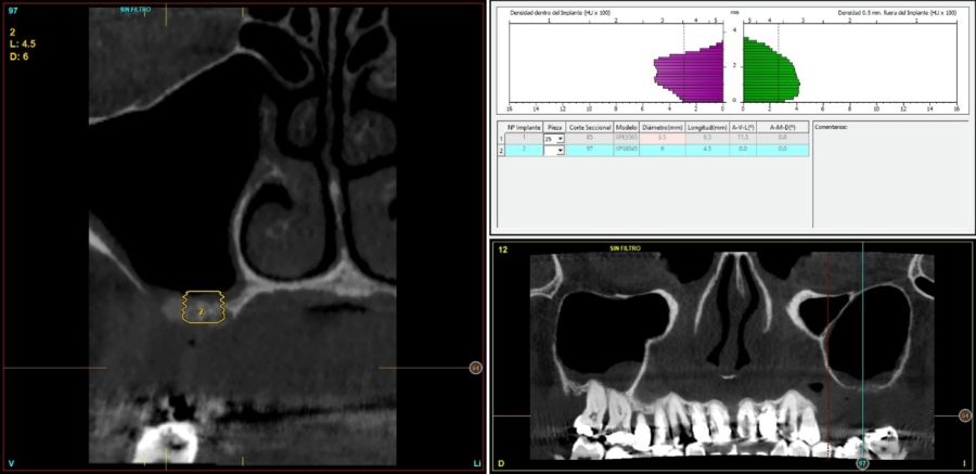
Patients were retrospectively included if, between June 2020 and September 2021, they had undergone placement of 4.5-mm-long dental implants with direct cortical anchorage in residual ridges up to 4.5 mm in height, together with transcrestal sinus floor elevation using autologous bone harvested during drilling and PRGF-Endoret as the graft material. Before implant placement, all patients were assessed using diagnostic models, an intraoral examination and a cone-beam computed tomography (CBCT) scan. The scans were analysed using specialised software (BTI-Scan III) to determine the exact residual ridge height and bone density and to adapt the surgical protocol and implant selection accordingly. As part of the preoperative protocol, patients received antibiotic premedication with amoxicillin (2 g orally) one hour before the procedure, together with paracetamol (1 g orally) as an analgesic. After surgery, patients continued antibiotic therapy with amoxicillin (500–750 mg orally every 8 hours, according to body weight) for five days. All implants were placed by the same surgeon using the biological drilling technique, characterised by low-speed drilling without irrigation. The drilling protocol was adapted to the density and volume of bone at the recipient site to ensure adequate primary implant stability8-9.
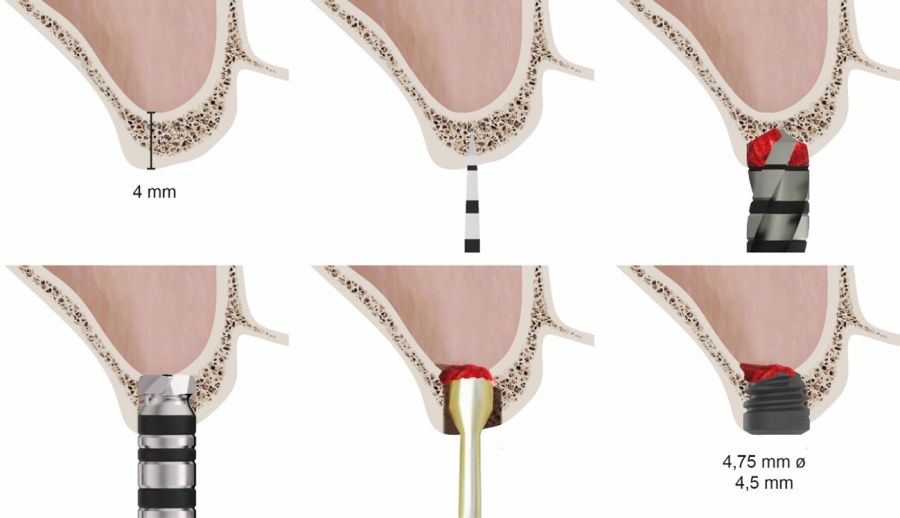
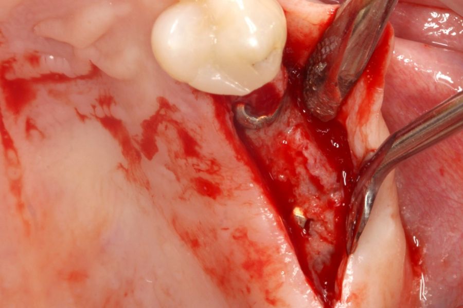
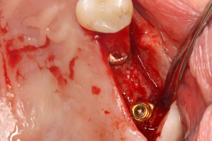
Final access to the cortical bone of the sinus floor was achieved with a drill specifically designed for this technique (a front-cutting drill), which allows controlled removal of the maxillary sinus floor without compromising the integrity of the Schneiderian membrane¹⁰. Once the membrane had been exposed through the crestal osteotomy, it was elevated in a controlled manner. The graft was then placed and consisted of autologous bone harvested during drilling at other implant sites prepared during the same surgical procedure and combined with PRGF-Endoret. The implant was placed using a surgical motor set to a maximum torque of 25 Ncm and a speed of 25 rpm. Final insertion was performed manually using a torque wrench to ensure optimal fixation (Figure 1).
Implant loading was performed four to five months later, initially with a provisional prosthesis under progressive loading, in all cases using transepithelial abutments. The definitive prosthesis was placed one to two months after initial loading.
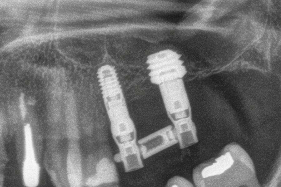
To estimate marginal bone loss, a reference of known length on the radiographs, specifically, the implant length, was used to calibrate the measurements. Following calibration, the software used (Digora for Windows; SOREDEX Digital Imaging Systems) calculated the actual measurements. Marginal crestal bone loss was determined by measuring the distance from the implant shoulder to the first bone-to-implant contact. To assess changes in bone resorption in each patient, the radiograph taken at the time of prosthesis placement was used as the reference. The patients subsequently attended follow-up visits every six months, during which new follow-up radiographs were obtained. During postoperative visits and after placement of the prosthesis, any prosthetic or surgical complications were recorded in the clinical record and subsequently analysed.
Statistical analysis
The primary outcome was implant survival, and the secondary outcome was crestal bone stability after loading. The patient was considered the unit of analysis for variables such as age, sex and medical history, whereas the implant was the unit of analysis for all other variables.
The Shapiro–Wilk test was used to assess the normality of the data distribution. Qualitative variables were analysed using frequency distributions, whereas quantitative variables were described using the mean and standard deviation.
The implant survival rate was estimated using the Kaplan–Meier method. All statistical analyses were performed using SPSS version 15.0 (SPSS Inc., Chicago, IL, USA), with a significance level of 5% (p < 0.05).
This study was designed as a retrospective study based on the review of clinical data previously recorded in routine practice. All procedures were performed in accordance with the ethical principles of the Declaration of Helsinki and applicable local regulations governing research involving human participants. Written informed consent was obtained from all included patients, authorising both the clinical treatment and the use of their data for scientific purposes. As no additional interventions were performed and no treatment plans were modified for research purposes, and data collection was limited to the retrospective analysis of cases, prior review by an institutional ethics committee was not considered necessary.
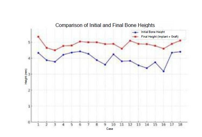
A total of 16 patients were included, in whom 18 implants meeting the previously described selection criteria were placed. The mean age of the patients was 58.76 years (±11.43), and 12 patients were women. The mean preoperative bone height at the implant sites was 3.96 mm (±0.38), with a range of 3.18–4.43 mm.
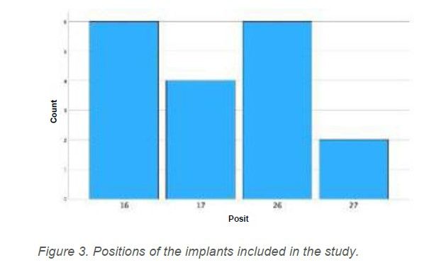
In all cases, the amount of sinus floor elevation ranged from 0.2 to 1.3 mm. Figure 2 shows the initial and final bone heights for each implant. The most frequent implant positions were 16 and 26, each accounting for 33.3%,whereas position 27 was the least frequent (11.1%). All implant positions are shown in Figure 3.
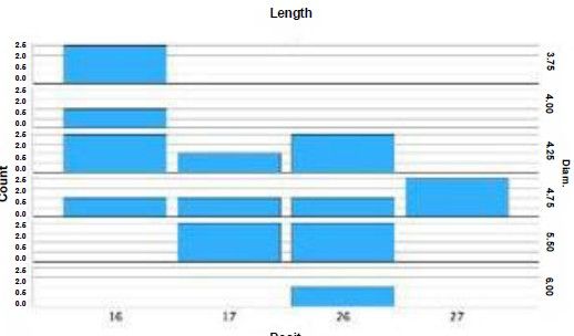
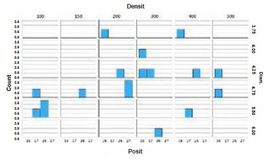
Implant diameters ranged from 3.75 to 6 mm. The most frequent diameters were 4.25 and 4.75 mm, each accounting for 27.8%. All implants placed were 4.5 mm long. The diameters and lengths of the implants included in the study according to their position are shown in Figure 4. Type IV bone was the most common bone type at the implant sites, occurring in 46.7% of cases, with a mean density of 263.89 HU (±134.82). Figure 5 shows the bone densities for all implants according to position and diameter.
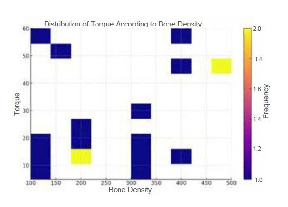
The implant insertion torque ranged from 5 Ncm to 60 Ncm, with a mean of 28.33 Ncm (±17.65). The most frequent insertion torque was 15 Ncm (27.8% of cases). Figure 6 shows the insertion torque values obtained according to bone density in Hounsfield units.
The mean follow-up period for the implants was 36.3 months (±2.71; range, 36–42 months). No implant failures occurred during this period, resulting in a cumulative survival rate of 100%. The mean mesial bone loss at the end of follow-up was 0.35 mm (±0.62) for all implants, while the mean distal bone loss was 0.11 mm (±0.49).
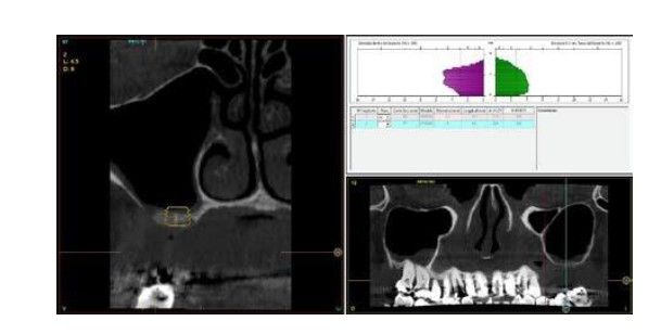
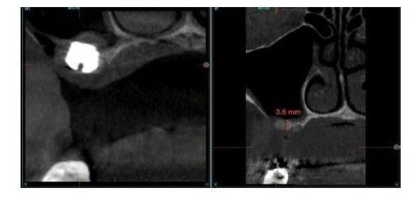
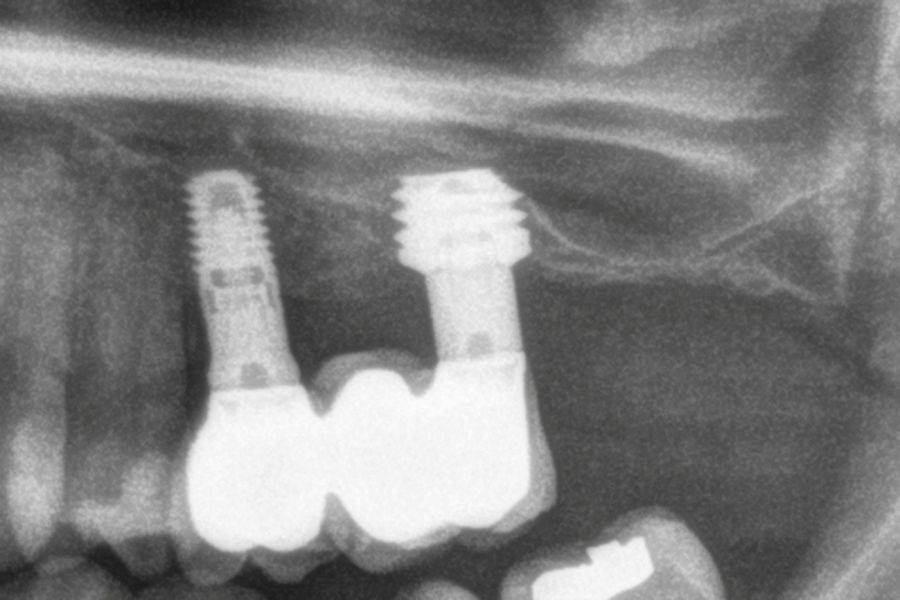
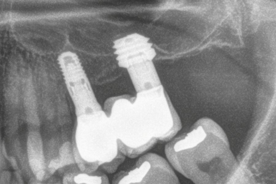
Figures 7–13 show one of the cases included in the study.
Implant dentistry is increasingly moving towards minimally invasive procedures, in line with other medical specialties. In this context, the use of smaller implants has become particularly important. In particular, 4.5-mm-long implants, such as those analysed in the present study, represent a viable therapeutic alternative for the rehabilitation of patients with moderate-to-severe bone atrophy in the posterior maxilla, with or without the need for transcrestal sinus floor elevation. This strategy can optimise functional outcomes and reduce the surgical morbidity associated with more invasive procedures, such as sinus floor elevation via a lateral approach11-15. Short and extra-short implants have also proved to be a predictable treatment option, as demonstrated by systematic reviews with meta-analyses reporting survival rates for extra-short implants ranging from 86.7% to 100%16,17. In particular, 4-mm-long implants have more recently been included in this category. Existing studies of their survival, crestal bone loss and surgical or prosthetic complications indicate outcomes comparable to those of conventional-length implants placed in conjunction with regenerative techniques17-19. However, regenerative techniques are generally associated with increased patient morbidity, reinforcing the importance of extra-short implants as a less invasive treatment option. The introduction of 4.5-mm-long implants, combined with an appropriate diagnostic and surgical protocol, has made it possible to optimise primary stability even at sites with very low bone density or in patients with minimal residual bone volume. This development has facilitated the use of transcrestal sinus floor elevation, allowing 4.5-mm-long implants to be placed at sites with a residual bone height of only 1–2 mm¹². As a result, the treatment approach to rehabilitation of the atrophic posterior maxilla has changed significantly, reducing the need for more invasive procedures and improving clinical outcomes. At sites with greater residual bone height, these implants can also be placed without the need for sinus access. This is an advantage, as transcrestal sinus floor elevation would have been necessary with 5.5-mm-long implants20-23.
According to the data obtained in the present study, 4.5-mm-long implants proved to be a predictable treatment option for rehabilitation of the posterior maxilla in cases of limited residual bone height when combined with transcrestal sinus floor elevation. These implants therefore represent a suitable treatment option in cases where residual bone height is between 4 and 5 mm.

Anitua, Eduardo
Private practice in oral implantology, Eduardo Anitua Foundation, Vitoria.
Clinical researcher, Eduardo Anitua Foundation, Vitoria, Spain. University Institute for Oral Regenerative Medicine and Implantology (UIRMI), Vitoria, Spain.
Correspondence:
Dr. Eduardo Anitua
Eduardo Anitua Foundation; C/ Jose
Maria Cagigal 19, 01007 Vitoria, Spain;
Phone: +34 945160653,
[email protected]