
Original article
Comparison of available bone retromolar space for maxillary molar distalization in adult patients with class I and class II malocclusions
Introduction: The distalization of upper molars is a conservative alternative in the treatment of class II malocclusion to achieve a class I dental relationship. The available bone space is measured from the distal root of the second upper molar to the vestibular cortical bone, rather than the distance from the crown of the second molar to the maxillary tuberosity.
Objective: To compare the available retromolar bone space for maxillary molar distalization in adult patients with class I and class II malocclusions.
Methods: A descriptive, observational, cross-sectional, and retrospective study. The sample consisted of 120 cases from the database of the Orthodontics Graduate Program at the Universidad Autónoma de Nayarit. The space was measured using a line parallel to the upper occlusal plane passing through the furcation of the second upper molar. Distances were taken from the most distal point of the distal root of the second upper molar to the inner edge of the maxillary cortical bone.
Results: Using a Student’s t-test, statistically significant differences were found in the retromolar space between patients with class I and class II. It was found that there was an association between retromolar space and sex and the SNA angle in class II.
Conclusions: There are significant differences in retromolar space between patients with class I and class II malocclusions, with higher values in class II. Sex and the SNA angle were found to influence retromolar space in patients with class II.
Keywords: Distalization; Malocclusion; Class I; Class II; Retromolar space.
The maxillary tuberosity (MT) is a bilateral anatomical structure located at the distal and inferior border of the infratemporal surface of the maxilla. The alveoli of the third molars are located in this region, which is closely related to other anatomical structures, including the pterygomaxillary fissure at its posterior boundary and the floor of the maxillary sinus at its superior boundary. These anatomical relationships give the maxillary tuberosity an important role in molar distalization procedures¹.
This anatomical arrangement facilitates an efficient biomechanical approach to molar distalization, as it allows en masse retraction of the maxillary dentition. Molar distalization may be performed bilaterally or unilaterally, particularly in cases of Class II malocclusion¹.
Maxillary molar distalization offers a means of avoiding dental extractions. Distalization mechanics are indicated for the treatment of patients with skeletal and dental Class II malocclusion. This procedure involves moving the molars distally to achieve Class I molar and canine relationships and relieve anterior crowding by creating space for dental alignment. It is commonly used when extractions are to be avoided, particularly in patients with compromised facial profiles²˒³.
Distalization is indicated in patients with erupted second molars close to the occlusal plane, increased overjet due to protrusion of the anterior teeth (maxillary or mandibular, depending on the malocclusion class), minimal skeletal discrepancies, a neutral or horizontal growth pattern, mild or moderate crowding, and in those who do not wish to undergo extractions⁴.
It is essential to consider the patient’s facial biotype and growth pattern. The available bone space must also be assessed during distalization. This space is measured from the distal root of the second molar to the buccal cortex, rather than from the crown of the second molar to the MT⁵.
Various options are available for gaining space in conjunction with distalization, including proclination of the anterior teeth, expansion, interproximal enamel reduction (stripping), extractions and orthognathic surgery. The choice of the most appropriate option depends on the severity of the case and the patient’s facial profile³.
Early molar distalization mechanics were associated with undesirable effects, such as anchorage loss in the premolars and incisors, which delayed treatment. Adverse effects were also observed in the maxillary first molar, including extrusion, tipping and distal rotation. Adverse effects of distalization mechanics include posterior rotation of the mandible, increased lower facial height, increased facial convexity and an increase in the mandibular plane angle. These effects are favourable in patients with a deep bite and those with a brachyfacial pattern, but unfavourable in patients with an open bite and those with a dolichofacial pattern⁶.
Several methods can be used to achieve molar distalization, including extraoral appliances (such as headgear) and intraoral appliances (such as the Pendulum appliance and spring-based orthodontic appliances). Patient cooperation is essential, particularly with extraoral mechanics, which led to the development of fixed intraoral appliances that deliver light, continuous forces⁴˒⁷.
Temporary skeletal anchorage techniques are currently used, including mini-implants inserted into bone and miniplates placed in the maxilla, palate and zygomatic bone. These advances help improve anchorage and accelerate distalization⁸.
Orthodontists must be familiar with the protocol for measuring the available bone space using cone-beam computed tomography (CBCT), as this facilitates treatment planning. This measurement is essential for determining whether molar distalization is a viable option and likely to be successful, and directly influences treatment duration and effectiveness.
The aim of the present study is to compare the available retromolar bone space for maxillary molar distalization in adult patients with Class I and Class II malocclusions.
This was a descriptive, observational, cross-sectional study. The amount of space available for maxillary distalization was assessed using CBCT scans from 120 patients in the database of the Postgraduate Programme in Orthodontics at the Universidad Autónoma de Nayarit. The patients had previously undergone CBCT scanning with a VATECH PaX-i3D unit for orthodontic indications. The participants were selected by non-probability sampling until 60 Class I and 60 Class II patients had been included.
The inclusion criteria were: all patients aged between 20 and 40 years with available CBCT scans, skeletal Class I or Class II malocclusion, complete dentition up to the second molars, an alveolar bone level coronal to the molar furcations and full patient records.
The exclusion criteria were previous orthodontic treatment, previous tooth extractions (except third molars), facial asymmetry (>3 mm according to Ricketts’ posteroanterior cephalometric analysis), dental prostheses and craniofacial syndromes.
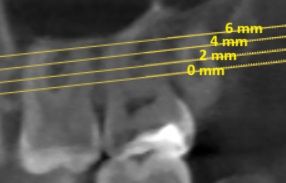
The maxillary occlusal plane was used as a reference for measuring the available bone space. This plane connects the mesiobuccal cusps of the maxillary first molars with the incisal edge of the maxillary right central incisor. An additional plane parallel to the maxillary occlusal plane and passing through the furcation of the maxillary second molar was designated the 0-mm plane. Three further parallel planes were defined 2, 4 and 6 mm apical to this plane (Figure 1).
At each of these levels, the cusp line was used as the reference for measuring the retromolar distance. The cusp line was defined as the line connecting the buccal cusps of the maxillary first and second molars.
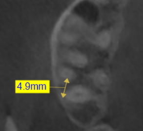
Linear distances were measured along the cusp line from the most distal point of the distal root of the right maxillary second molar to the inner border of the buccal cortical plate of the MT (Figure 2).
Cephalometric analyses were performed by manual tracing on acetate sheets using a light box to identify and measure the relevant cephalometric landmarks and planes:
SNA: Angle used to assess the anteroposterior position of the maxilla relative to the cranial base. It is formed by the Sella–Nasion (S–N) and Nasion–Point A (N–A) lines.
SNB: Angle used to assess the anteroposterior position of the mandible relative to the cranial base. It is formed by the Sella–Nasion (S–N) and Nasion–Point B (N–B) lines.
ANB: Angular difference between the N–A and N–B lines. It is used to assess the sagittal relationship between the maxilla and the mandible.
IMPA: Angle formed between the long axis of the mandibular incisor and the mandibular plane (Go–Gn). It assesses the inclination of the mandibular incisor relative to the mandible.
U1-SN: Angle between the long axis of the maxillary incisor and the S–N plane. It is used to assess the inclination of the maxillary incisor relative to the cranial base.
U1/ANS–PNS: Angle between the long axis of the maxillary incisor and the palatal plane formed by the anterior nasal spine and posterior nasal spine (ANS–PNS). It reflects the inclination of the maxillary incisor relative to the hard palate.
The data and descriptive statistics were tabulated in a Microsoft Excel spreadsheet. Student’s t-test and analyses of variance (ANOVA) were performed using StatCalc version 8.1.3.
This study was approved by the Research Ethics Committee of the Academic Unit of Dentistry at the
Universidad Autónoma de Nayarit (approval number UAO/CEI/038/2025).
The mean age was 24.52 ± 5.05 years in the Class I group and 24.04 ± 5.54 years in the Class II group. The following mean values were found in the skeletal Class I group: the ANB angle was 2.76° ± 1.11°; retromolar space was 3.95 ± 1.89 mm at 0 mm, 4.4 ± 2.01 mm at 2 mm, 4.70 ± 1.98 mm at 4 mm and 4.94 ± 2.35 mm at 6 mm. The descriptive statistics for all the variables are in Table 1.
In the skeletal Class II group, the mean ANB angle and retromolar measurements were as follows: ANB angle, 6.54° ± 1.54°; retromolar space, 5.08 ± 1.90 mm at 0 mm, 5.55 ± 1.91 mm at 2 mm, 5.65 ± 2.02 mm at 4 mm and 5.53 ± 2.05 mm at 6 mm (Table 1).
Age, sex and the SNA, SNB, ANB, IMPA, U1/SN and U1/ANS–PNS angles were assessed by linear regression to determine whether they were associated with retromolar space. In the Class I group, none of these variables was associated with retromolar space. In patients with Class II malocclusion, retromolar space was associated with both sex and the SNA angle, with greater values in men and in those with a higher SNA angle (Table 2).
Student’s t-test was used to compare the variables between the Class I and Class II groups. Statistically significant differences were found for all variables except the angle between the maxillary central incisor and the Sella–Nasion plane and the angle between the maxillary central incisor and the palatal plane (ANS–PNS) (Table 1).
The space available for molar distalization has been assessed in different studies, each using specific methods and approaches to evaluate retromolar space. Hui et al.⁹ measured the available space in the maxilla using six levels based on plane 0, with 1.5-mm increments distally, and three additional planes extending apically at 3-mm intervals. The results ranged from 3.89 to 4.30 mm, with a mean available distalization space of 4.06 mm in patients with Class II malocclusion. This type of measurement is relevant because it reflects the amount of space available for molar movement, a crucial factor in planning orthodontic distalization treatment.
Yilmaz et al.¹⁰, on the other hand, analysed maxillary retromolar space using lateral cephalometric radiographs, measuring from the line marking the anterior border of the mandibular ramus to the posterior border of the maxillary second molar. The study found a mean retromolar space of 11.3 mm and observed that this space was smaller in women than in men. This sex-related difference in the amount of space suggests that anatomical characteristics may influence the success of distalization treatment.
Ye et al.¹¹ compared the space available for maxillary distalization using two methods: panoramic radiographs with cephalometric landmarks and CT images with anatomical boundaries. On the three-dimensional images, the mean sagittal distance from the posterior nasal spine (PNS) to the posterior border of the maxillary tuberosity was 1.80 ± 1.81 mm, whereas the shortest distance between the distal border of the maxillary tuberosity and the most distal root of the maxillary second molar was 5.24 ± 2.26 mm. In addition, the cephalometric distance from the distal height of contour of the maxillary second molar to the PNS was 5.98 ± 2.62 mm. These results highlight the importance of using three-dimensional imaging to assess the available space more accurately.
Choi et al.¹² compared the available mandibular space on 110 CBCT scans from patients with Class I and Class III malocclusions. The results showed that the available space was greater in patients with Class III malocclusion. In this group, the mean distance along the posterior line of occlusion was 6.0 ± 3.3 mm at the furcation level and decreased to 2.7 ± 2.8 mm as the measurement was taken closer to the root apex. This decreasing pattern suggests that patients with Class III malocclusion have less space for molar distalization closer to the root apex.
Regarding mandibular retromolar space, previous studies based on panoramic radiographs assumed that the anterior border of the mandibular ramus was the posterior anatomical limit for molar distalization. However, recent studies have shown that the true limit is the inner cortical plate of the mandible because of the cortical resistance encountered when the root reaches the inner border of the buccal cortical plate¹³.
Following this criterion, the maxillary limit could be considered the distance from the distal root of the maxillary second molar to the inner border of the buccal cortical plate of the MT. This underscores the importance of considering bone anatomy when planning distalization treatment.
Distalization involves moving the molars towards the MT; therefore, the amount of distalization depends on the volume of bone available in the direction of root movement. The success of this procedure is closely related to the topography of the MT, which may vary between patients.
In this regard, López et al.¹ analysed 277 MTs and found that both width and height were smaller in the absence of the third molar. This suggests that the presence of the third molar may influence the space available for distalization and should therefore be considered during treatment planning.
In the present study, patients with Class II malocclusion had greater retromolar bone space than those with Class I malocclusion at all the levels assessed (0, 2, 4 and 6 mm). This finding suggests that molar distalization planning may be more favourable in individuals with Class II malocclusion, increasing the feasibility of non-extraction treatment.
In the regression analysis, sex and the SNA angle were significantly associated with retromolar space in the Class II group. Male sex and a larger SNA angle were correlated with greater available space, which could be explained by anatomical differences in the size and shape of the maxilla between men and women.
One of the main limitations of the study was the age range of the patients (20–40 years), which limits the generalisability of the results to growing adolescents and older adults. In addition, third-molar status was not considered, although this factor may influence the space available for distalization.
Finally, it is recommended that the anatomical limit for molar distalization be determined using CBCT imaging, as this provides more accurate predictions than conventional radiography.
2D radiographs may be affected by errors arising from incorrect image orientation, superimposition of structures and magnification distortion, which could compromise measurement accuracy and treatment success.
Statistically significant differences in retromolar space were observed between patients with Class I and Class II malocclusions, with greater retromolar space values in patients with Class II malocclusion. This finding suggests that the space available for molar distalization may be more favourable in patients with Class II malocclusion, which could influence clinical decision-making when planning orthodontic treatment according to the type of malocclusion. Sex and the SNA angle were found to influence retromolar space in patients with skeletal Class II malocclusion.


Baeza-Rivera, Jatziri
Dental surgeon Universidad Autónoma de Nayarit, Mexico. Student in the Orthodontics Specialty Programme at the Universidad Autónoma de Nayarit, Mexico.
Silva-Zatarain, Aida Nabile
Dental surgeon, Universidad Autónoma de Nayarit, Mexico. Master’s degree in Dental Sciences (Orthodontics), Benemérita Universidad Autónoma de Puebla, Mexico. Lecturer in the Orthodontics Specialty Programme at the Universidad Autónoma de Nayarit, Mexico.
Gutiérrez-Rojo, Jaime Fabián
Dental surgeon, Universidad de Guadalajara. Specialist in Orthodontics, Universidad Autónoma de Nayarit, Mexico. Master’s degree in Public Health, Universidad Autónoma de Nayarit, Mexico. Lecturer in the Orthodontics Specialty Programme at the Universidad Autónoma de Nayarit, Mexico.
Correspondence:
Jatziri Baeza Rivera.
Unidad Académica de Odontología
Universidad Autónoma de Nayarit Ciudad de la Cultura S/N, CP 63155
Tepic, Nayarit (México) [email protected]