Ir al contenido principal
Publicación

ESPECIAL EN INGLÉS. Científica Dental vol. 19 2022

Año: 2022
Número: 4
Volumen: 19

Clinical case

Published in spanish Científica Dental Vol. 19. Nº 2. 2022 www.cientificadental.es

Block grafting in combination with the two-stage split technique for the rehabilitation of a case of severe bone resorption of the maxilla in width

Resumen

The use of different surgical techniques to achieve the resolution of the most complex atrophy cases is of vital importance.
Increasingly, we have cases with edentulism of longer duration in our practice that require implant treatment with fixed prosthesis, which is a challenge when planning and carrying out treatments. To do this, we can use different techniques that allow us to gain width and height, as well as implants of different lengths and diameters that allow us to adapt to each situation. In the present clinical case, we show a rehabilitation that combines different surgical techniques to achieve the desired result.

Abstract
Este artículo está exclusivamente disponible para su descarga en PDF. | 03/23/2024
Palabras clave
Block grafting, Dental implant, Split technique
Introducción

More and more frequently, patients with long-term edentulism, wearing complete prosthesis come for dental consultation requesting implantology treatment. The digital age, social networks and patient access to clinical information in dentistry, means that the latest advances in rehabilitation of extremely atrophic maxillary and mandible reach the patients and they demand a solution to their situation, they thought it could not be corrected in any other way than with removable dentures1-4. Therefore, patients sometimes request implantology treatments in cases of great complexity that make us have to use all the surgical and prosthetic tools at our disposal to solve them5.

When we face a severe resorption in a horizontal sense, both in the maxilla and in the mandible, there are different techniques that help us to recover the lost bone volume to later insert dental implants6-7. Each of the techniques has its indications and may, sometimes, in the same patient, need several of these techniques to achieve a regeneration of the lost bone volume, since there are substantial variations in the quality and quantity of bone volume within the same arch of the same patient, as well as areas where in addition to width the bone plates have been completely or partially lost, which changes the approach of the technique to employ6-9.
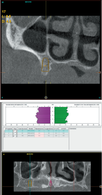
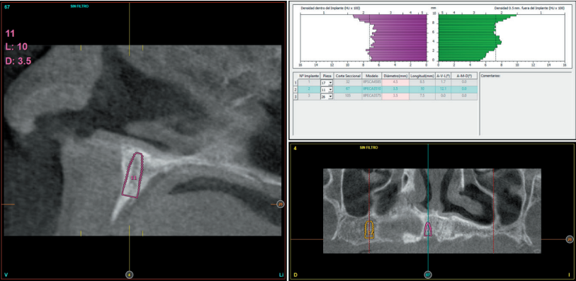
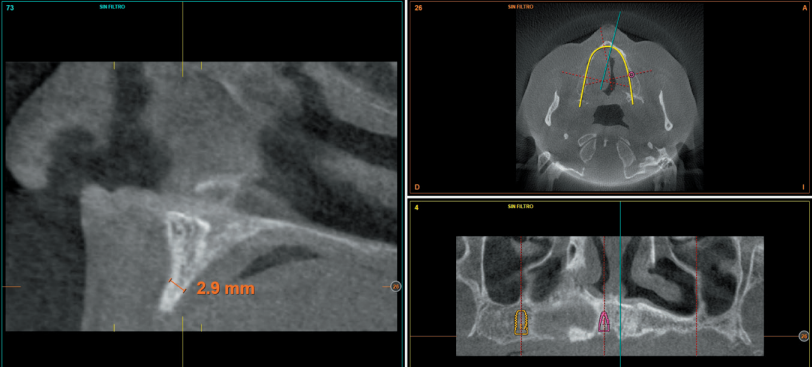
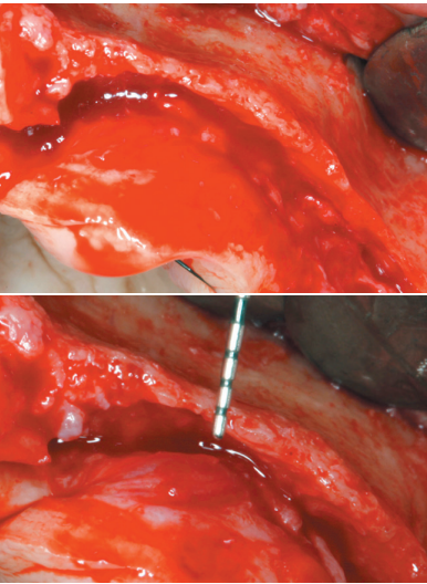
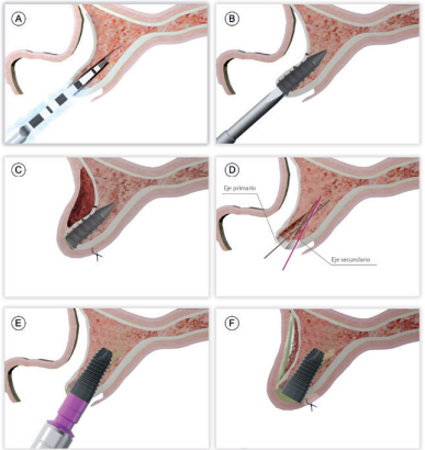
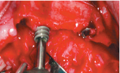
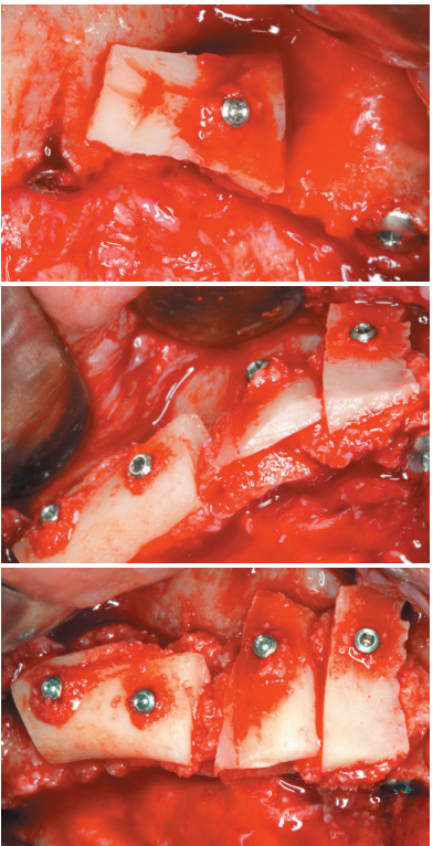
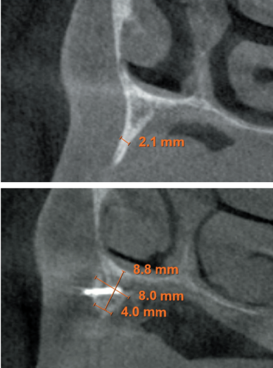
In general, when there is a reduced bone width (below 3 mm of residual crest) that does not allow direct insertion of the implant we can opt for an expansion or split crest, in one or two phases, as long as the two bone plates are available and there is a bone particulate between them that allows the separation between them by inserting the definitive implant or the expander implant10-14. When one of the two bone plates is absent, in that case, the recommendation will be to use block grafts or guided bone regenerations13-14.
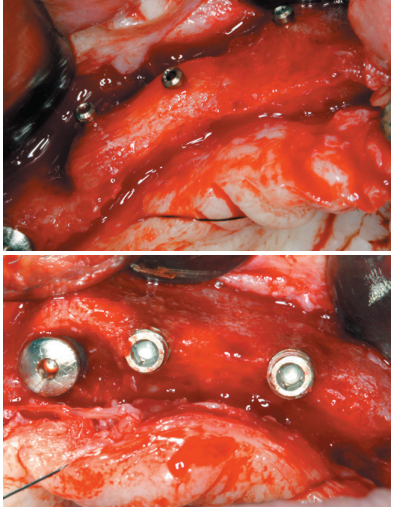
Both techniques are widely documented15, and the selection between one procedure and the other is mainly based on the availability of autologous bone to perform block surgery, and the ability of the operator to perform the technique, since the use of blocks requires a greater learning curve and can lead to more complications in obtaining, manipulating and healing, since it is a technique of greater technical difficulty.
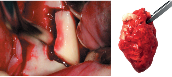
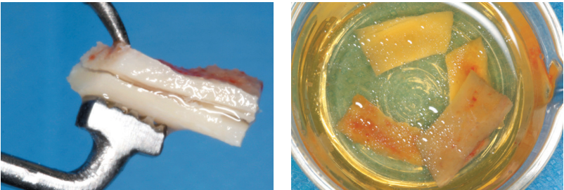
In spite of all this, if careful protocols are used to obtain the graft, and a careful positioning technique is performed, paying special attention to soft tissues, nowadays, it can be performed without greater rate of complications than the bone regeneration technique, especially when the intraoral area is used as the donor zone and the obtained bone is maximized by dividing the block as described by Khoury16-18.
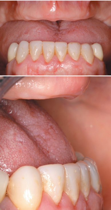
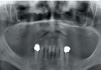
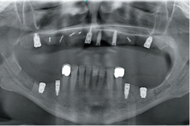
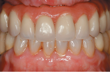
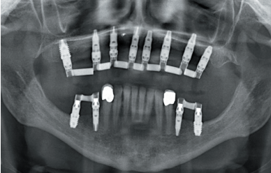
In the following clinical case, a situation is shown where different procedures have been necessary to rehabilitate the maxilla with extreme bone loss in width, depending on each of the areas to be treated, individualizing the type of treatment according to the characteristics of the remaining bone recess.

Bibliografía
1

Kashbour WA, Rousseau NS, Thomason JM, Ellis JS. Provision of information on dental implant treatment: Patients’ thoughts and experiences. Clin Oral Implants Res 2018;29(3):309-19.

2

Apfel PS. Wisdom. N Y State Dent J
2015;81(5):10-3

3

Elfadil S, Johnston B, Normand C, Allen F, O’Connell B. An investigation of the characteristics of edentulous patients who choose or refuse implant treatment. Int J Prosthodont 2021;34(2):147–153

4

Kakudate N, Yokoyama Y, Sumida F,
Matsumoto Y, Riley JL 3rd, Gordan VV, Gilbert GH. Practice-based research agenda priorities selected by patients: findings from a dental practicebased research network. Int Dent J 2019;69(3):183-91.

5

De Groot RJ, Oomens MAEM, Forouzanfar T, Schulten EAJM. Bone
augmentation followed by implant surgery in the edentulous mandible: A systematic review. J Oral Rehabil 2018;45(4):334-43.

6

Tolstunov L, Hamrick JFE, Broumand V, Shilo D, Rachmiel A. Bone augmentation techniques for horizontal and vertical alveolar ridge deficiency in oral implantology. Oral Maxillofac Surg Clin North Am 2019;31(2):163-91.

7

Elnayef B, Porta C, Suárez-López Del
Amo F, Mordini L, Gargallo-Albiol J,
Hernández-Alfaro F. The fate of lateral ridge augmentation: A systematic review and meta-analysis. Int J Oral Maxillofac Implants 2018;33(3):622-35.

8

Esposito M, Grusovin MG, Felice P,
Karatzopoulos G, Worthington HV, Coulthard P. The efficacy of horizontal and vertical bone augmentation procedures for dental implants – a Cochrane
systematic review. Eur J Oral Implantol 2009;2(3):167-84.

9

Sanz-Sánchez I, Ortiz-Vigón A, SanzMartín I, Figuero E, Sanz M. Effectiveness of lateral bone augmentation on the
alveolar crest dimension: A systematic review and meta-analysis. J Dent Res 2015;94(9 Suppl):128S-42S

10

Cortese A, Pantaleo G, Amato M, Claudio PP. Ridge expansion by flapless split crest and immediate implant placement: Evolution of the technique. J Craniofac Surg 2016;27(2):e123-8.

11

Waechter J, Leite FR, Nascimento
GG, Carmo Filho LC, Faot F. The split crest technique and dental implants: a systematic review and meta-analysis. Int J Oral Maxillofac Surg 2017;46(1):116-28

12

Anitua E, Begoña L, Orive G. Clinical
evaluation of split-crest technique with ultrasonic bone surgery for narrow ridge expansion: status of soft and hard tissues and implant success. Clin Implant Dent Relat Res 2013;15(2):176-87.

13

Anitua E, Alkhraisat MH. Is Alveolar
Ridge Split a Risk Factor for Implant Survival? J Oral Maxillofac Surg 2016;74(11):2182-91.

14

Anitua E, Begoña L, Orive G. Two-stage split-crest technique with ultrasonic bone surgery for controlled ridge expansion: a novel modified technique. Oral Surg Oral Med Oral Pathol Oral Radiol Endod
2011;112(6):708-10.

15

Mendoza-Azpur G, de la Fuente A,
Chavez E, Valdivia E, Khouly I. Horizontal ridge augmentation with guided bone regeneration using particulate xenogenic bone substitutes with or without
autogenous block grafts: A randomized controlled trial. Clin Implant Dent Relat Res 2019;21(4):521-30.

16

Chappuis V, Cavusoglu Y, Buser D, von Arx T. Lateral ridge Augmentation using autogenous block grafts and guided bone regeneration: A 10-year prospective case series study. Clin Implant Dent Relat Res 2017;19(1):85-96.

17

Aghaloo TL, Moy PK. Which hard tissue augmentation techniques are the most successful in furnishing bony support for implant placement? Int J Oral Maxillofac Implants 2007; 22 Suppl:49-70. Erratum in: Int J Oral Maxillofac Implants 2008;
23(1):56.

18

Sánchez-Sánchez J, Pickert FN,
Sánchez-Labrador L, GF Tresguerres
F, Martínez-González JM, Meniz-García C. Horizontal ridge augmentation: A comparison between Khoury and Urban technique. Biology (Basel). 2021 5;10(8):749.

19

Anitua E, Carda C, Andia I. A novel drilling procedure and subsequent bone autograft preparation: a technical note. Int J Oral Maxillofac Implants. 2007; 22(1):138-45. Erratum in: Int J Oral Maxillofac Implants 2007; 22(2):309.

20

Khoury F, Hanser T. Mandibular bone
block harvesting from the retromolar
region: a 10-year prospective clinical study. Int J Oral Maxillofac Implants 2015; 30(3):688-97.

21

Khoury F. Mandibular bone block grafts: Diagnosis, instrumentation, harvesting, technical and surgical procedures. In Khoury F AH, Missika P (ed) Bone Augmentation in Oral Implantology, (ed. Chicago, Quintessence, 2007, p 169).

22

Solakoglu Ö, Heydecke G, Amiri N, Anitua E. The use of plasma rich in growth factors (PRGF) in guided tissue regeneration and guided bone regeneration. A review of histological, immunohistochemical, histomorphometrical, radiological and clinical results in humans. Ann Anat 2020; 231:151528.

23

Chou TM, Chang HP, Wang JC.
Autologous platelet concentrates in
maxillofacial regenerative therapy.
Kaohsiung J Med Sci 2020; 36(5):305-10.

24

Anitua E, Prado R, Orive G. Platelet-rich plasma therapies: Building the path to evidence. J Orthop 2016;14(1):68-9

25

Anitua E, Sánchez M, Zalduendo MM,
de la Fuente M, Prado R, Orive G, Andía I. Fibroblastic response to treatment with different preparations rich in growth factors. Cell Prolif 2009; 42(2):162-70.

26

Anitua E, Begoña L, Orive G. Controlled ridge expansion using a two-stage splitcrest technique with ultrasonic bone surgery. Implant Dent 2012;21(3):163-70.

27

Bare-Welchel B, Campbell PM, Gonzalez M, Buschang PH. Effects of bone grafting, performed with corticotomies and buccal tooth movements, on dehiscence formation in dogs. Am J Orthod Dentofacial Orthop 2017;151(6):1034-47.

28

Esposito M, Thomsen P, Ericson LE,
Lekholm U. Histopathologic observations on early oral implant failures. Int J Oral Maxillofac Implants 1999;14(6):798-810.