Artículo original
Implantes cortos y extracortos en maxilar y mandíbula con bajo torque de inserción. Estudio retrospectivo con un seguimiento de 1 a 7 años
Introducción: La realización de implantes cortos y extracortos en situaciones de extrema reabsorción maxilar y mandibular supone que la colocación del implante se realiza en condiciones de escaso volumen óseo residual y, por lo tanto. puede ser posible que el torque de inserción que se logre sea menor que en otras situaciones. En el presente artículo se investiga la posible relación que puede tener este bajo torque de inserción con la supervivencia del implante y la pérdida ósea crestal.
Material y método: Fueron revisadas historias clínicas de forma retrospectiva de los implantes insertados en el segundo semestre del año 2015 y primer trimestre del año 2021, para identificar implantes cortos y extracortos con torque de inserción < 20 N/cm. El implante fue la unidad de análisis para la estadística descriptiva en cuanto a la localización, dimensiones del implante, y mediciones radiográficas. El paciente fue la unidad de medida para el análisis de la edad, sexo y la historia médica. Las variables cualitativas se describieron mediante un análisis de frecuencias y las cuantitativas mediante media y desviación estándar. La supervivencia delos implantes se calculó mediante el método de Kaplan-Meier.
Resultados: Fueron reclutados 36 pacientes en los que se insertaron 40 implantes que cumplieron los criterios de inclusión marcados para el estudio. El torque de inserción de los implantes reclutados varió entre 5 y 15 Ncm, siendo 5 Ncm en el 37,5% de los casos, 10 Ncm en el 30% de los casos y 15 Ncm en el 32,5 % restante. El tiempo medio de seguimiento de los implantes estudiados fue de 75 meses (+/- 35; rango 12-
84 meses). Durante el tiempo de seguimiento no existió ningún fracaso de los implantes estudiados, registrándose únicamente dos complicaciones por rotura de provisional y que representaron el 5% de la muestra. La pérdida ósea media mesial registrada para el conjunto de los implantes fue de 0,63 mm (+/- 0,52) y la media de la pérdida ósea distal fue de 0,48 mm (+/-0,64).
Conclusiones: El torque de inserción bajo, en situaciones límite de volumen óseo residual o de densidad ósea, no genera una mayor tasa de fracaso en los implantes estudiados, siempre que se siga un protocolo conservador con el lecho óseo receptor y se individualice en cada caso en función de las características del implante a insertar y del volumen óseo remanente.
Introduction: The use of short, extrashort implants in situations of extreme maxillary and mandibular resorption means that the implant is placed in conditions of low residual bone volume and therefore it is possible that the insertion torque achieved may be lower than in other situations. This article investigates the possible relationship that this low insertion torque may have with implant survival and crestal bone loss.
Methods: Medical records of implants inserted in the second half of 2015 and first quarter of 2021 were retrospectively reviewed to identify short, extra-short implants with insertion torque < 20 N/cm. The implant was the unit of analysis for descriptive statistics in terms of location, implant dimensions, and radiographic measurements. The patient was the unit of measurement for the analysis of age, sex and medical history. Qualitative variables were described by frequency analysis and quantitative variables by mean and standard deviation. Implant survival was calculated using the Kaplan-Meier method.
Results: Thirty-six patients were recruited and 40 implants were inserted that met the inclusion criteria for the study. The insertion torque of the recruited implant ranged from 5 to 15 Ncm, with 5 Ncm in 37.5% of cases, 10 Ncm in 30% of cases and 15 Ncm in the remaining 32.5%. The mean follow-up time of the implants studied was 75 months (+/- 35; range 12-84 months). During the follow-up time there was no failure of the implants studied, with only two complications due to temporary (resin) breakage being recorded and which represent a total of 5% of the sample. The mean mesial bone loss recorded for all implants was 0.63mm (+/- 0.52) and the mean distal bone loss was 0.48mm (+/-0.64).
Conclusions: Low insertion torque, in borderline situations of residual bone volume or bone density does not generate a higher failure rate in the implants studied, provided that a conservative protocol is followed with the recipient bone bed and is individualised in each case according to the characteristics of the implant to be inserted and the remaining bone volume.
Los implantes cortos y extracortos se insertan en la mayoría de las ocasiones en situaciones anatómicas límite, donde existe un escaso volumen óseo residual de forma directa o unidos a técnicas rehabilitadoras (crecimiento vertical, elevación transcrestal)1,2. Por ello, en muchas de estas situaciones lograr una correcta estabilidad primaria del implante es más complicado que con implantes de mayor longitud y lechos óseos con mayor volumen. Para lograr una estabilidad primaria suficiente, que evite los micromovimientos durante la fase de integración del implante y asegure su viabilidad en la fase más temprana del seguimiento, se han ideado protocolos de fresado para distintos lechos óseos, adaptándose el fresado (tanto en número de fresas como en profundidad) a las características del lecho receptor y a la morfología del implante3-5. Estas secuencias de fresado individualizadas nos aseguran una correcta estabilidad inicial en la mayoría de los casos, incluso cuando existe un volumen óseo escaso o una baja densidad y los implantes son además cortos o extra-cortos9-11. Según la literatura internacional, para que la integración sea correcta tras la inserción del implante, no deben existir micromovimientos superiores a los rangos entre 50 y 150 μm. Cuando se superan estos rangos, se puede generar una fibrointegración con el consecuente fracaso de la oseointegración15,16. Es difícil calcular el torque de inserción necesario para que no se generen este tipo de micromovimientos
perjudiciales para la integración, encontrándose en algunos estudios cifras que rondan los 20 Ncm, aunque también existen trabajos de investigación que demuestran que implantes con un torque de inserción <20Ncm pueden producir tasas de supervivencia favorables y un mantenimiento óptimo de los niveles óseos marginales, incluso después de la carga funcional inmediata17,18. Por ello, se ha decidido analizar la evolución de forma retrospectiva de una cohorte de implantes con un torque de inserción por debajo de 20 Ncm, en diferentes localizaciones y situaciones clínicas, con el fin de conocer el comportamiento en cuanto a la integración, supervivencia a largo plazo y pérdidas óseas crestales.
Fueron revisadas historias clínicas de forma retrospectiva de los implantes insertados en el segundo semestre del año 2015 y primer trimestre del año 2021, para identificar aquellos casos implantes cortos y extra-cortos (BTI- Biotechnology institute, superficie Unicca®) insertados en zona de atrofia ósea tanto mandibular como maxilar con bajo volumen óseo residual y torque de inserción final menor a 20 Ncm. Todos los pacientes fueron estudiados antes de la inserción de los implantes mediante modelos diagnósticos, exploración intraoral y realización de un TAC dental (Cone-Beam), analizado posteriormente mediante un software específico (BTI-Scan II). Antes de la inserción de los implantes se utilizó una premedicación antibiótica consistente en amoxicilina 2gr vía oral una hora antes de la intervención y paracetamol 1 gramo vía oral (como analgésico). Posteriormente los pacientes prosiguieron con un tratamiento de amoxicilina 500-750 mg vía oral cada 8 horas (según peso) durante 5 días. Los implantes fueron colocados por un mismo cirujano, mediante la técnica de fresado biológico, a bajas revoluciones, sin irrigación1,2,9,11. Los pacientes acudieron posteriormente para la realización de radiografías panorámicas de control y sobre estas radiografías se realizaronn las mediciones necesarias para comprobar la estabilidad y pérdida ósea crestal de los implantes. Una vez obtenida la radiografía en formato digital se calibró mediante un software específico (Digora for Windows, SOREDEX Digital Imaging Systems) a través de una longitud conocida en la radiografía como es el implante dental. La recolección de los datos fue llevada a cabo por dos examinadores independientes. Análisis estadístico El implante fue la unidad de análisis para la estadística descriptiva en cuanto a la localización, dimensiones del implante, y mediciones radiográficas. El paciente fue la unidad de medida para el análisis de la edad, sexo y la historia médica. Fue realizado un test de Shapiro-Wilk sobre los datos obtenidos para constatar la distribución normal de la muestra. Las variables cualitativas se describieron mediante un análisis de frecuencias y las cuantitativas mediante media y desviación estándar. La supervivencia de los implantes se calculó mediante el método de Kaplan-Meier. Los datos fueron analizados con SPSS v15.0 para Windows (SPSS Inc., Chicago, IL, USA).
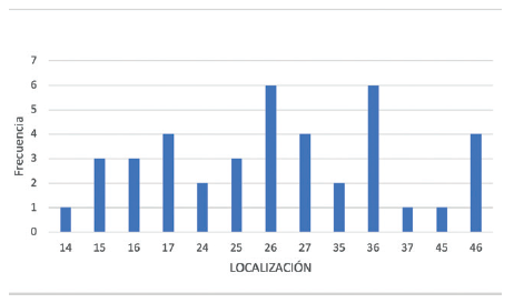
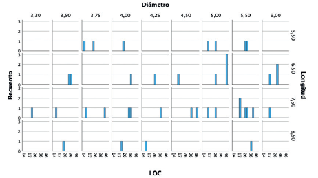
Fueron reclutados 36 pacientes en los que se insertaron 40 implantes que cumplieron los criterios de inclusión marcados para el estudio. El 65% de los pacientes reclutados fueron de sexo femenino, con una media total de la muestra de 55,7 años (+/- 13,98). El torque de inserción de los implantes reclutados varió entre 5 y 15 Ncm, siendo 5 Ncm en el 37,5% de los casos, 10 Ncm en el 30% de los casos y 15 Ncm en el 32,5 % restante. El tipo óseo en la zona de inserción de los implantes fue de tipo IV en el 67,5% de los casos y tipo III en el 32,5% restante. La localización de los implantes mayoritaria fue para la zona de 36 y 26 (15% para ambos casos), seguido de las posiciones 17, 27 y 46 con un (10% de los casos respectivamente). El resto de las posiciones incluidas en el estudio se muestran en la Figura 1. La longitud de los implantes estudiados osciló entre 5,5 y 8,5 mm, siendo la más frecuente la de 7,5 (42,5%). El diámetro osciló entre 3,30 mm y 6 mm, siendo el más frecuente el de 5 y 5,5 mm (20% respectivamente). El resto de los diámetros y longitudes incluidos en el estudio se muestran en la Figura 2. El tiempo medio de seguimiento de los implantes estudiados fue de 75 meses (+/- 35; rango 12-84 meses). Los implantes se insertaron de forma directa en el 65% de los casos, realizándose elevación de seno transalveolar en el 12,5% de los casos y elevación transalveolar con sobrecorrección en el 5% de los casos. En el 17,5 % de los pacientes se llevó a cabo sobrecorrección vestibular como única técnica accesoria. Todos los implantes se rehabilitaron en dos fases quirúrgicas, realizándose una prótesis provisional de carga progresiva de resina con estructura de barras articuladas en todos los casos, tras la segunda fase, y se rehabilitaron ferulizados a otros implantes tanto en la fase de provisionalización como en la fase de prótesis definitiva. Durante el tiempo de seguimiento no existió ningún fracaso de los implantes estudiados, registrándose únicamente dos complicaciones por rotura de provisional (resina), que no dificultó que el provisional siguiera cumpliendo su función, y que representó un total del 5% de la muestra. La pérdida ósea media mesial registrada para el conjunto de los implantes fue de 0,63 mm (+/- 0,52) y la media de la pérdida ósea distal fue de 0,48 mm (+/-0,64).
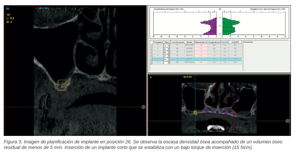
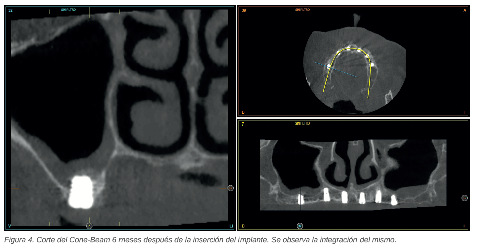
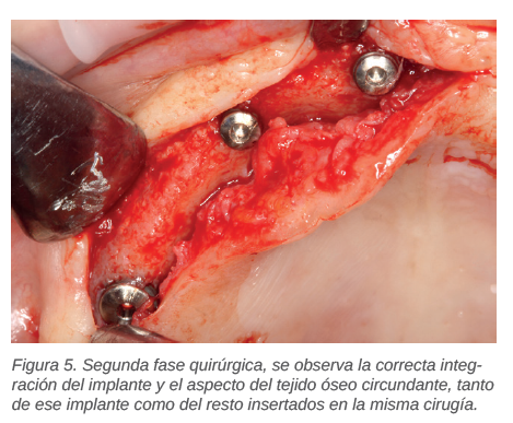
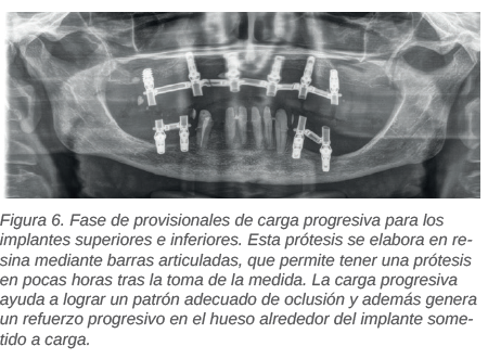
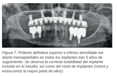
En las Figuras 3-7 se muestra uno de los casos incluidos en el estudio.
El principal reto al que nos enfrentamos cuando rehabilitamos zonas tanto de maxilar como de mandíbula con extrema reabsorción o con una densidad ósea muy baja es la consecución de estabilidad primaria suficiente para la correcta oseointegración de los implantes, sobre todo, cuando los implantes empleados son cortos o extracortos y además se emplean técnicas accesorias. 10-19-20. En estos casos extremos, tanto por volumen de lecho óseo como por densidad o por la unión de ambos, lograr la estabilidad primaria adecuada depende principalmente de adaptar de forma precisa la secuencia de fresado al lecho receptor, teniendo en cuenta por supuesto las características derivadas de la geometría (macro y micro) del implante empleado21-23. En el estudio llevado a cabo por Santamaría-Arrieta y cols.24 se pone de manifiesto como la realización de la preparación del neoalveolo buscando una compresión que nos aporte estabilidad inicial mejora los torques de inserción, en situaciones donde no es fácil conseguir la estabilidad primaria. Dentro de que la compresión nos puede garantizar la estabilidad, se debe tener además en cuenta que una compresión excesiva podría generar una inflamación elevada con alta liberación de citoquinas, y ser
contraproducente en cuanto a lograr un mejor anclaje del hueso a largo plazo25-27. Por ello, un protocolo de fresado cuidadoso adaptado al lecho receptor en función de las características del mismo es de crucial importancia y puede garantizar mejores resultados, tal como se muestra en el presente artículo, donde no han existido fracasos a pesar de situarse los implantes en diferentes localizaciones, con escaso volumen óseo residual o escasa densidad, incluso empleándose técnicas quirúrgicas accesorias. En la literatura internacional, existen trabajos en los que se pone de manifiesto que un bajo torque de inserción no influye en la supervivencia de los implantes a medio plazo, englobándose en la mayoría de los trabajos torques por debajo de 20 Ncm, como los reportados en el presente estudio17,8,28. En estos trabajos se pone énfasis también en la preparación del alveolo como hecho crucial en garantizar la oseointegración del implante.
El torque de inserción bajo, en situaciones límite de volumen óseo residual o de densidad ósea, no genera una mayor tasa de fracaso en los implantes estudiados, siempre que se siga un protocolo conservador con el lecho óseo receptor y se individualice en cada caso, en función de las características del implante a insertar y del volumen óseo remanente.

Anitua, Eduardo
DDS, MD, PhD. Práctica privada
en implantología oral, Clínica
Eduardo Anitua, Vitoria, España.
Instituto universitario para la
medicina oral regenerativa y
la implantología – (University
Institute for Regenerative
Medicine and Oral Implantology
– UIRMI) (UPV/ EHU Fundación
Eduardo Anitua), Vitoria, España.
BTI Biotechnology institute (BTI),
Vitoria, España.
Correspondencia:
Dr. Eduardo Anitua
[email protected]
Fundación Eduardo Anitua C/ Jose
Maria Cagigal 19, 01007, Vitoria.
Indexada en / Indexed in:
– IME
– IBECS
– LATINDEX
– GOOGLE ACADÉMICO